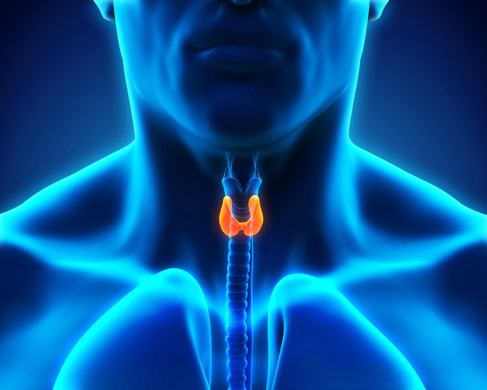
Primary hyperparathyroidism is a common disorder in which one or more parathyroid glands are abnormally enlarged. Surgical removal of the enlarged glands is the definitive treatment. The traditional surgical approach of four-gland exploration has been largely replaced with minimally invasive parathyroidectomy (MIP), which reduces the incidence of such associated complications as permanent hypocalcemia and recurrent laryngeal nerve injury. The success of the minimally invasive technique lies in a good preoperative localizing study to identify the diseased gland. This enables the surgeon to make a small incision to remove the diseased gland, thus obviating the need to look at other glands and minimizing the risk of complications.
At Roswell Park Comprehensive Cancer Center, our study of choice is a four-dimensional computerized tomography (4DCT) with volume rendering reconstruction to identify the abnormal parathyroid glands. This technique has largely replaced the routine use of sestamibi SPECT technology. In our experience, based on 200 consecutive parathyroidectomies, 4DCT was positive in 96% of cases as compared to 65.4% with sestamibi SPECT and 57.7 % with ultrasound in patients with single-gland disease, demonstrating superior sensitivity with 4DCT1. In addition, 4DCT avoided unnecessary opposite-side exploration in 32% of patients when compared with sestamibi in patients with single-gland disease.
Because cost and radiation exposure are primary concerns with 4DCT, we have modified the 4DCT technique to reduce radiation exposure and selectively eliminate routine intraoperative parathyroid hormone (PTH) testing. To minimize radiation exposure, we utilize a lower tube current for non-contrast and delayed images, giving an average radiation dose of 11–13 mSv2. The most recent implementation of ASIR technology has further reduced the radiation exposure and approaches the range of sestamibi as well as essentially 50% of that previously reported in the literature for 4DCT.
Avoiding Intraoperative PTH Monitoring
Furthermore, intraoperative PTH testing is routinely employed with MIP. Using our prospective database, we have devised a scientifically derived algorithmic index, utilizing biochemical and imaging characteristics, which enables us to preoperatively predict which patients have single-gland disease, with a 100% positive predictive value3. Therefore, we can avoid the routine use of intraoperative PTH monitoring in these patients, reducing both operating time and time under anesthesia, and realizing a significant cost reduction as well. We are currently validating this index in an investigational trial.
As evidenced, we have made — and continue to make — important advances in the management of this disease. In our opinion, using 4DCT as a preoperative localizing study combined with selective elimination of intraoperative PTH testing is the safest, most cost-effective strategy for treating this endocrine disorder.
Learn more about Endocrine Surgery for Hyperparathyroidism at Roswell Park.
References
Kukar M, Platz TA, Schaffner TJ , Elmarzouky R, Groman A, Kumar S, Abdelhalim A, Cance WG. The Use of Modified Four-Dimensional Computed Tomography in Patients with Primary Hyperparathyroidism: An Argument for the Abandonment of Routine Sestamibi Single-Position Emission Computed Tomography (SPECT). Ann Surg Oncol. 2015 Jan; 22(1):139-45.
Platz TA, Kukar M, Elmarzouky R, Cance WG, Abdelhalim A. Low-dose four-dimensional CT with volume rendering reconstructions for primary hyperparathyroidism: How I do it. World J Radiol. 2014 Sep 28; 6(9):726-9.
Kukar M, Cho E, Platz TA, Attwood K, Abdelhalim A, Kumar S, Cance WG. Development of an index to predict single-gland parathyroid disease and selectively eliminate intraoperative parathyroid hormone testing. 67th Annual Society of Surgical Oncology Meeting, March 12–15, 2014, Phoenix, AZ.

