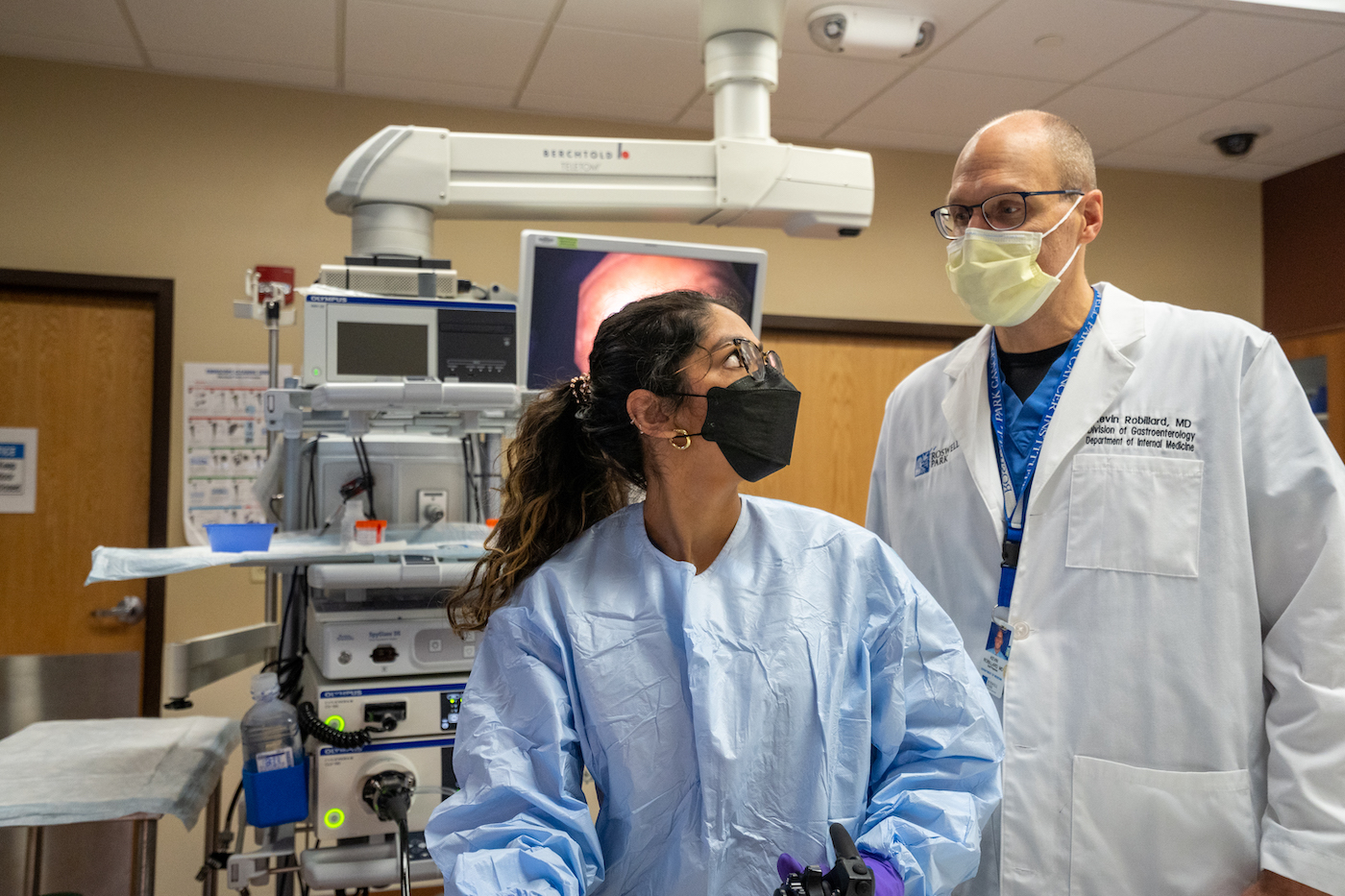
Endoscopy is a minimally invasive procedure to examine the inside of the gastrointestinal tract using an endoscope, a thin flexible tube with a light and camera. No surgical incisions are made to insert the endoscope, which is instead introduced through either the mouth or the anus.
Roswell Park Comprehensive Cancer Center’s Endoscopy Center combines leading-edge technology with minimally invasive techniques to diagnose and treat malignant and benign digestive conditions. Many of the specialized endoscopic procedures offered at Roswell Park are not available at other centers in the region.
Services we offer
An endoscope is passed into the rectum and advanced throughout the colon. During the procedure, the doctor slowly withdraws the endoscope, examining for pre-cancerous and cancerous polyps or growths. These polyps can be removed at the time of identification, thereby preventing cancer. This procedure requires a thorough colon cleanse beforehand to ensure a high-quality examination and proper visualization.
Like a colonoscopy, an endoscope is passed into the patient, however this time via the mouth. The upper gastrointestinal tract, including the esophagus, stomach, and duodenum are examined for any abnormalities.
A wireless ‘camera pill’ is used to look at the inside of the small intestines and other parts of the digestive tract. The small intestines, on average, are greater than 20 feet long. The patient swallows a capsule about the size of a large pill, which contains a tiny wireless camera that takes pictures inside the body as it travels through the digestive tract and sends them to a small recorder worn on the patient’s waist or shoulder. The pictures are put together to make a movie of the intestine. The capsule passes out of the body during a bowel movement and does not need to be retrieved.
This procedure uses a specialized endoscope and an overtube with balloons attached. The balloons are inflated and deflated to allow the physician to pass the endoscope deeper into the small intestine than what can be achieved with a standard endoscope. This procedure may help some patients avoid surgery and is typically used when other procedures are unable to reach difficult areas of the small intestine.
Like an upper endoscopy, an endoscope is passed through the patient's mouth and stomach into the first part of the small intestine. However, at the tip of the endoscope is an ultrasound device. The doctor slowly withdraws the endoscope from the intestine toward the stomach to take images of the pancreas and surrounding organs and tissues, including the liver, bile ducts, gallbladder, and lymph nodes.
Using an ultrasound device inside the intestines, the doctor can often better evaluate these areas compared to an ultrasound probe placed on the skin. A needle biopsy may be taken during this procedure, allowing for a minimally invasive and painless means to obtain tissue samples.
This is a procedure to diagnose and treat problems in the liver, gallbladder, bile ducts and pancreas. After an endoscope is passed through the patient's mouth, stomach, and into the first part of the small intestine, a smaller tube (catheter) is passed through the endoscope and into the bile ducts and pancreatic ducts. Dye is injected through the catheter to the duct, and x-rays then guide the physician to allow them to obtain samples, remove bile duct stones or place a stent to relieve an obstruction.
Electric shocks are delivered in short pulses to break up large bile duct stones during an endoscopic retrograde cholangiopancreatogrphy (ERCP) procedure.
An endoscopic procedure where a camera is inserted into the ducts of the liver and pancreas to evaluate, treat or take biopsies during an endoscopic retrograde cholangiopancreatography (ERCP) procedure.
A procedure that uses radio waves to heat and destroy abnormal cells, such those that occur with Barrett’s esophagus. The radio waves travel through electrodes (small devices that carry electricity). Radiofrequency ablation can prevent progression to cancer and also be used to treat other conditions.
A procedure that uses liquid nitrogen to freeze abnormal tissues such as Barrett’s esophagus or esophageal cancer. The technology flash-freezes water in cells causing cell death in the abnormal tissue.
A procedure where a solution is injected through an IV that makes tumor cells sensitive to light. The endoscope is then used to shine light at the tumor causing the tumor cells to decay.
A procedure that uses an endoscope to place a metal stent into the intestines of the upper or lower digestive tract to relieve a blockage. Stent placement results in expansion of the narrowed intestines and improve symptoms of blockage.
A procedure that places a flexible tube into the stomach through the abdominal wall to provide nutrition and liquids to a patient who is unable to take enough food or liquids by mouth.
A procedure used to treat recurrent infections of the colon that are not responsive to standard antibiotics, namely C. Difficile, or infections that do not go away or keep coming back. Healthy bacteria are transferred from a donor without infection to the patient’s colon, usually via colonoscopy. Then the new healthy bacteria from the donor are able to suppress the unhealthy bacteria causing the infection.
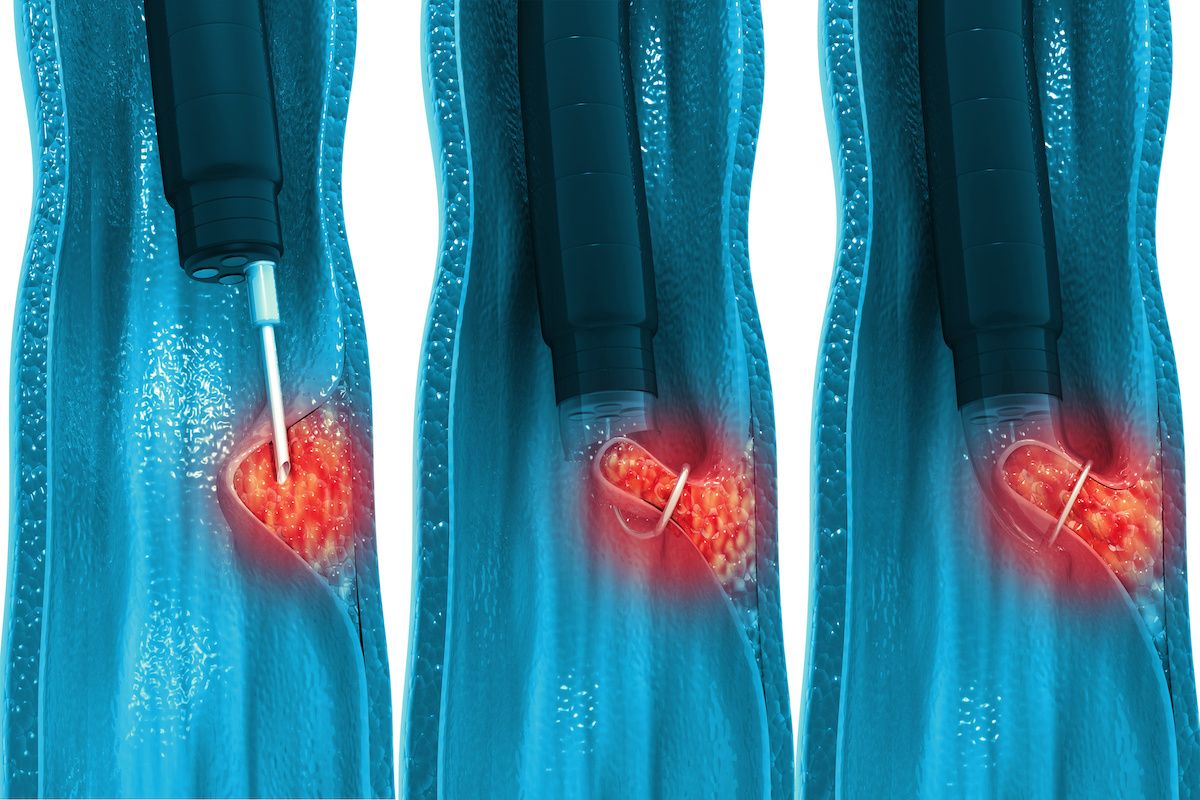
Endoscopic mucosal resection (EMR) and endoscopic submucosal dissection (ESD) are procedures to remove cancerous or other abnormal tissues (lesions) from the digestive tract, such as large polyps, precancerous tissue, or superficial early cancers. Fluid is injected to raise the tissue after which the tissue is removed using a combination of tools introduced through the endoscope. The goal is to remove the superficial tissue of the bowel but leave the deeper tissue. (Surgery would typically remove all of the layers of tissue.) This provides a minimally invasive approach to remove abnormal tissue, thereby avoiding a more involved surgery.
An ultrasound scope is used to drain pancreatic fluid collections into the gastrointestinal tract with placement of a stent between the collection and the intestines, allowing a path of drainage.
Patients who have had previous gastric bypass surgery are often unable to have standard endoscopic retrograde cholangiopancreatography (ERCP) to access the bile ducts. This procedure uses the ultrasound endoscope to place a stent between the stomach pouch to the excluded part of the stomach, creating a passageway between them. The scope is then driven through the stent to perform an ERCP.
Anatomy altered by previous surgery and/or malignancy can often make standard endoscopic retrograde cholangiopancreatography (ERCP) impossible. The ultrasound endoscope can provide access to the bile duct/pancreatic duct to allow drainage and avoid the need for more invasive procedures.
Conditions we treat
- Cancer screening (colon, esophageal, gastric, small bowel and pancreatic)
- Tumors of the esophagus, stomach, small bowel, colon, rectum, liver, bile duct, gallbladder and pancreas (malignant and benign)
- Strictures (narrowing) of the esophagus, duodenum, and colon, including post-surgical strictures
- Barrett’s esophagus
- Polyps, obstructions and/or bleeding of the gastrointestinal tract
- Bile duct stricture and stones
- Pancreatitis, pancreatic cysts and pseudocysts
- Inherited polyposis syndromes, such as Familial Adenomatous Polyposis (FAP)
- Recurrent Clostridioides difficile (C Difficile) infection
Endoscopy Center at Roswell Park
Our center brings together Endoscopy and Interventional Pulmonology services into one state-of-the-art facility for diagnosis and treatment of malignant and benign gastrointestinal and pulmonary conditions.
With our unique capabilities, we offer minimally invasive options in place of traditional surgery whenever possible, caring for patients in a way that affords a quicker recovery, less pain and fewer side effects. More than 90 percent of procedures are performed on an outpatient basis.
The Roswell Park advantage
- One of the few facilities in the region to offer many of these advanced endoscopic and pulmonary procedures.
- High-volume center for endoscopic ultrasound, ERCP, and bronchoscopy.
- Multidisciplinary teams involving Endoscopy, Surgery, Oncology, and Radiology allows for seamless and efficient coordination of care.


