Brain mapping and awake craniotomy
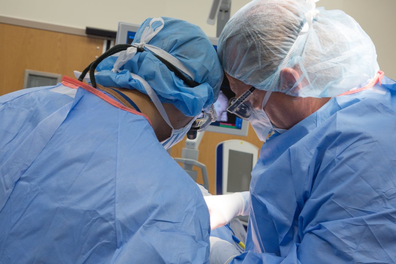
Because areas of the brain that control speech and movement look very similar to other parts of the brain, the neurosurgeon must be cautious when performing surgery in these areas in order to protect them. Our neurosurgeons use a technique called electrocortical mapping to provide the highest level of safety during brain tumor removal.
If it is necessary to monitor the patient’s language or motor function during surgery, the patient may remain awake during surgery but does not feel any pain, because the brain has no pain receptors. The patient looks at a series of images displayed on a computer monitor and answers questions while the neurosurgeon stimulates the surface of the brain with a very mild electrical current. The patient’s responses help the surgeon determine which areas of the brain control important functions, such as speech, so those areas can be avoided if possible.
Endoscopic skull base surgery
At Roswell Park, tumors located at the base of the skull are removed through expanded endoscopic endonasal surgery. The surgical team includes both a neurosurgeon and a head and neck surgeon working together. The head and neck surgeon inserts an endoscope — a thin, lighted telescope with a camera on one end — through one of the patient’s nostrils, and the camera sends images from inside the head to a flat-screen TV in the operating room. Surgical instruments are inserted through the other nostril so the head and neck surgeon can create a channel from the nose and sinuses to the skull base while viewing the surgical field on the TV. The neurosurgeon then removes the tumor using similar techniques.
This procedure is used to treat pituitary tumors, including those in patients with acromegaly and Cushing’s Disease, and for patients with meningiomas, chordomas, craniopharyngiomas, esthesioneuroblastomas and other midline skull base tumors.
Laser Interstitial Thermal Therapy (LITT)
Laser Interstitial Thermal Therapy, or LITT (brand name Visualase®), is a type of minimally invasive neurosurgery that uses heat to ablate (destroy) cancerous or other damaged tissue in the brain. Through a tiny incision (less than 2 cm), a laser probe delivers light energy to the area of concern, heating and destroying the abnormal tissue. The procedure is performed with MRI guidance so the neurosurgeon can monitor the temperature of the target area and receive real-time confirmation that all the necessary tissue has been destroyed and that the normal brain has been protected. The treatment does not require a large incision. Patients typically return home the day after the procedure.
LITT may be an option for patients with primary brain cancers, gliomas or metastatic brain tumors; for cancer that has returned after radiation or Gamma Knife therapy; and to treat such noncancerous conditions as epilepsy. LITT may also be used to destroy areas of radiation necrosis (swelling or edema (fluid buildup) caused by prior radiation therapy) in the brain.
LITT is available only at select centers across the nation.