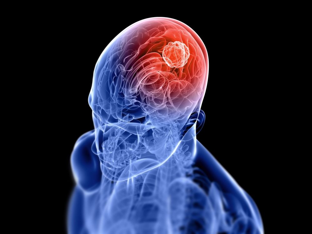
The words glioma and glioblastoma may look alike, and even sound alike, but the truth is more complicated.
“Glioma is a large category that includes a number of different types of tumors,” explains Lindsay Lipinski, MD, FAANS, Assistant Professor of Neurosurgery and Oncology at Roswell Park Comprehensive Cancer Center. The word “glia” means glue and glial cells are the ones that hold everything together in the brain by providing a framework to support and nourish the actual nerve cells which are also known as neurons.
Gliomas are graded on a scale of one to four. Grade one gliomas usually grow slowly and frequently behave in a more benign fashion. Grade two and grade three gliomas can grow more quickly and they frequently require more aggressive treatment. Grade four gliomas are the most aggressive type and are also known as glioblastoma. These tumors used to be called glioblastoma multiforme, or GBM for short.
“Lower grade gliomas typically occur in younger patients,” Dr. Lipinski says. “They often present with seizures and less commonly with neurological deficits such as paralysis or speech problems.” Sometimes lower-grade gliomas (grades one and two) can be found incidentally in patients who have an MRI or CT scan performed for a different reason, such as for migraines or minor head injury.
Most gliomas will require a biopsy to determine their grade and to predict their future behavior and growth rate. Grade one gliomas may be treatable with surgery alone and then monitored for regrowth without further treatment. However, grades two and three gliomas almost always require more aggressive treatment, which may include some combination of surgery, radiation therapy and chemotherapy. These gliomas can recur and can progress to become glioblastoma over time.
“Generally, the treatment for glioblastoma must be more aggressive because we know that its behavior involves fairly rapid growth. Watching and waiting doesn’t play a role,” Dr. Lipinski says. “We want a tissue diagnosis as soon as possible, either through a biopsy or removal of the tumor, and then chemotherapy and radiation must start pretty shortly after that.”
Identifying and typing a glioma
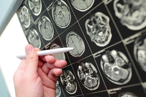
“Gliomas are typically seen well on a brain MRI scan,” Dr. Lipinski says, with MRI scans being much better than CT scans for seeing these tumors clearly.
In addition to the one-to-four grading system, gliomas are also classified based on the type of glial cell from which they develop. Glial cells that give rise to tumors in the brain can include ependymal cells (ependymomas), astrocytes (astrocytomas) and oligodendrocytes (oligodendrogliomas). Ependymal cells line the brain’s ventricles where spinal fluid is contained. Astrocytes provide the structural framework of the brain. Oligodendrocytes create the myelin sheath for the neurons that acts a little like insulation for wires throughout the brain’s communication system. Oligodendrogliomas frequently have a better prognosis, even at higher grades, and people diagnosed with oligodendroglioma tend to have a longer survival rate than patients with astrocytomas.
“Most gliomas are locally invasive but it is very uncommon for any glioma to metastasize outside of the nervous system to other organs,” Dr. Lipinski says. It’s also uncommon for gliomas to run in families, but “We know that people with gliomas may have a family history of other cancers, more so than patients who don’t have gliomas.”
Why Roswell Park for brain tumor treatment?
Brain tumors are very complex and require expert care. Learn what makes Roswell Park unique in diagnosing and treating brain tumors.
Learn MoreDon’t jump to conclusions about headaches
Dr. Lipinski stresses that headaches are rarely an indication of a brain tumor and that other causes like migraine are far more common.
“There are no real screening tests for glioma. For most patients, diagnosis is reliant upon someone having neurologic symptoms,” Dr. Lipinski says. “That could include having a seizure or developing paralysis, speech problems, partial loss of vision or impaired thinking. Patients who present with seizures tend to be younger and have less aggressive tumors. Lots of times, people with brain tumors come to an emergency room with stroke-like symptoms but it’s only when you get a scan that you realize it’s a tumor, not a stroke.”
Anyone diagnosed with a glioma is encouraged to follow the advice of their physician and understand that “not every situation is an emergency,” she adds. “There’s often time to get a second opinion.”