
Arriving at a diagnosis of bladder cancer may involve one or more of these steps:
- Urine tests to detect blood, cancer cells and other signs of disease. A 24-hour urine collection can measure volume of urine output, an indicator of kidney function.
- Bladder wash during which a solution is delivered through a catheter (tube) to the bladder to loosen cells on the bladder’s inner lining. The solution is removed, and a sample is examined under a microscope.
- Blood tests to assess kidney and liver function
- Cystoscopy to see inside the bladder and identify any tumor
- Imaging Scans to determine if a cancer has spread
- Transurethral Resection of Bladder Tumor (TURBT) to obtain a biopsy, confirm a cancer diagnosis, and determine the stage and grade of a cancer
It matters – getting the right diagnosis
Second opinion game-changer
If you seek a second opinion at Roswell Park, our bladder cancer team will review your tests, scans and pathology. In about 10% of cases we review, the diagnosis is changed, impacting your treatment options and decisions.
Getting an accurate diagnosis of your bladder cancer – including your cancer’s type, grade, shape, stage and whether it’s invasive or noninvasive – is essential to determining your treatment options and the way forward. At Roswell Park, our team of urologists, pathologists and radiologists, are uniquely experienced and specialize in performing and interpreting the tests, scans and procedures necessary for an accurate bladder cancer diagnosis.
What is a cystoscopy?
If you have symptoms of bladder cancer that cannot be attributed to another cause, such as an infection, your next step will be to undergo a simple procedure called flexible cystoscopy to allow your doctor to see the inside of your bladder. You may need a cystoscopy if:
- You have visible blood in your urine (urine may be rusty, pink or red)
- A urine test showed microscopic blood in your urine (not visible to your eyes)
- You have symptoms such as urinary frequency or urgency
In this procedure, a tube called a cystoscope (fitted with a tiny light and camera) is passed through the urethra and into your bladder, allowing your doctor to visualize the inside of your bladder and detect any tumors. This 15- to 30-minute procedure can be done in your doctor’s office and does not require anesthesia. A numbing gel is inserted into the urethra to prevent any discomfort. During this test, the doctor can collect cells from your bladder’s lining to examine, diagnose and even treat various bladder conditions, and perform a bladder wash.
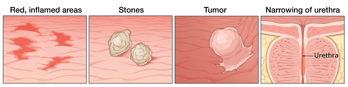
What does the doctor look for during cystoscopy?
Your doctor will look for any abnormalities in the urethra and lining of your entire bladder, such as:
Imaging scans — has my cancer spread?
If your flexible cystoscopy revealed bladder cancer, your medical team will need to determine whether or not the cancer has spread beyond your bladder, using one or more of the following imaging tests:
- Computerized Tomography (CT)
- Intravenous pyelography (IVP)
- Magnetic Resonance Imaging (MRI)
- Positron Emission Tomography (PET)
- Ultrasound
- Bone Scan
Although these scans can provide your medical team with a picture of your body’s structure and function and direct your medical team to an area of concern, scans cannot tell what is going on with your cells. A biopsy or tissue sample is needed to allow a pathologist to examine the cells and determine definitely whether it is cancer and which type.
What is Transurethral Resection of Bladder Tumor (TURBT)?
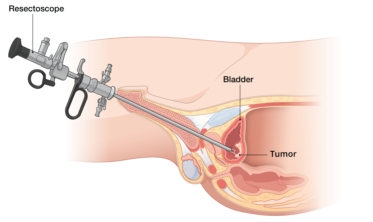
If your cystoscopy or imaging scans found a suspicious mass, tumor or any irregularity such as a lesion, your next step is to obtain a biopsy or tissue sample through a surgical procedure called Transurethral Resection of Bladder Tumor (TURBT). During a TURBT procedure, the surgeon inserts a tool called a resectoscope through the urethra to reach the inside of your bladder and remove a piece of tumor tissue, or the entire tumor from your bladder.