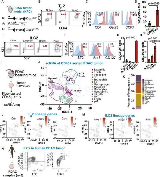
Immuno-phenotyping of pancreatic cancer revealed a dramatic expansion of TH2 (Cd4+Gata3+Ccr4+) cells within the CD4+ T cell population in the PDAC TME (72.1%) compared with the normal pancreas (8.33%) and spleen (0.4%).
This was accompanied by a significant increase in ILC2 cells (Lin−Sca1+ST2+) in the PDAC TME (74.2%) compared with the normal pancreas (7.96%), spleen (0.18%), and bone marrow (0.25%).
Specifically, the frequency of ILC2 cells was approximately 60% of lineage-negative (Lin−) cells in the PDAC (KPC model) compared with <10% in the normal pancreas and ∼25% in PanIN.
Similarly, the frequency of ILC2 cells was approximately 45% of Lin− cells in the PDAC (iKPC model) compared with <10% in the normal pancreas. Further, single-cell RNA sequencing (scRNA-seq) analysis identified the presence of the type 2 immunocytes in the PDAC TME.
Using previously reported gene signatures for TH2 and ILC2, we found that the TH2 cluster was enriched for Cd4, Gata3, and Ccr4 genes and ILC2 clusters were enriched for Hes1, Hs3st1, and Il1rl1 genes, which are bona fide markers of TH2 and ILC2 cells, respectively.
Finally, we analyzed fresh human PDAC samples by flow cytometry and found that ILC2 cells accounted for 14.2% of the Lin− cells. Overall, these results show that the murine and human PDAC TME contain abundant TH2 and ILC2s cells.
Contact the Dey Lab
Email: Prasenjit.Dey@RoswellPark.org
Phone: 716-845-1300, x5269
Office location: Center for Genetics & Pharmacology (CGP) L5-307
Lab Location: Center for Genetics & Pharmacology (CGP) L15-115
Department of Immunology
Roswell Park Comprehensive Cancer Center
Elm and Carlton Streets
Buffalo, NY 14263