Lighting up cancer cells with non-radioactive heavy water
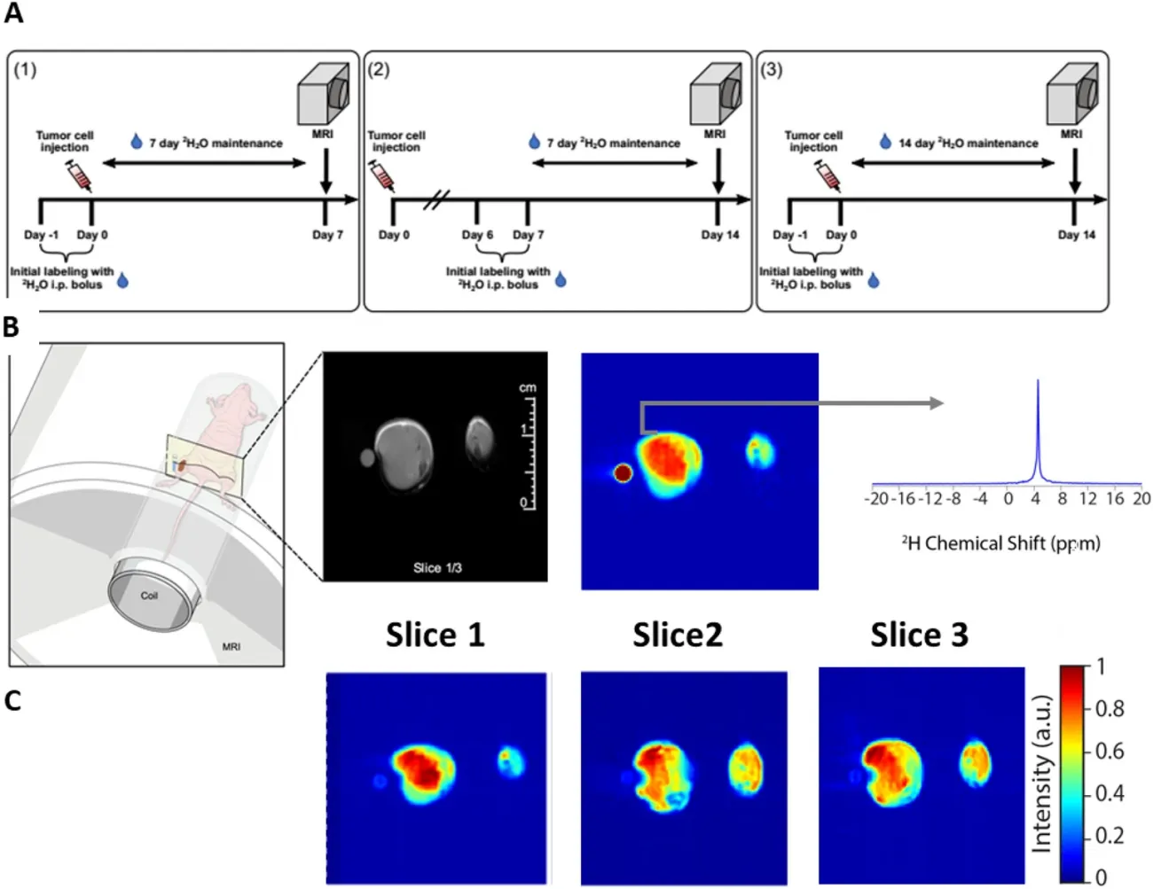
Deuterated water (2H2O) is a naturally occurring form of heavy water that is not radioactive. 2H2O looks and tastes like regular water (1H2O) but has a distinct signal from 1H2O on magnetic resonance imaging (MRI). Water is used by proliferating cells to make essential molecules that sustain high cell division rates, e.g., nucleic acids, proteins, metabolites, and lipids.
Cells that proliferate rapidly, such as cancer cells, become preferentially labeled with deuterium after short periods of systemic 2H2O administration and “light up” on deuterium magnetic resonance imaging (dMRI). For example, an individual with cancer can drink deuterated water and their tumor will “light up” on dMRI.
Detecting small tumors without radiation
Dr. Buxbaum’s team tested this imaging approach in several animal models and confirmed that small tumors are easily discernable from surrounding healthy tissues when using this approach. This method has broad clinical relevance and implications.
For example, dMRI may allow sensitive detection of small tumors without the use of radioactive isotopes or ionizing radiation, in contrast to positron emission tomography combined with computerized tomography (PET-CT), an imaging approach that is widely used in oncology.
Additionally, stable water isotopes may slow down proliferation of cancer cells. Therapeutic approaches combining the former with currently approved drugs are being explored by the Buxbaum laboratory.
An investigator-initiated clinical trial led by Dr. Buxbaum will soon open at Roswell Park to test deuterium MRI in lymphoma patients.
Deuterium imaging for CAR T cells
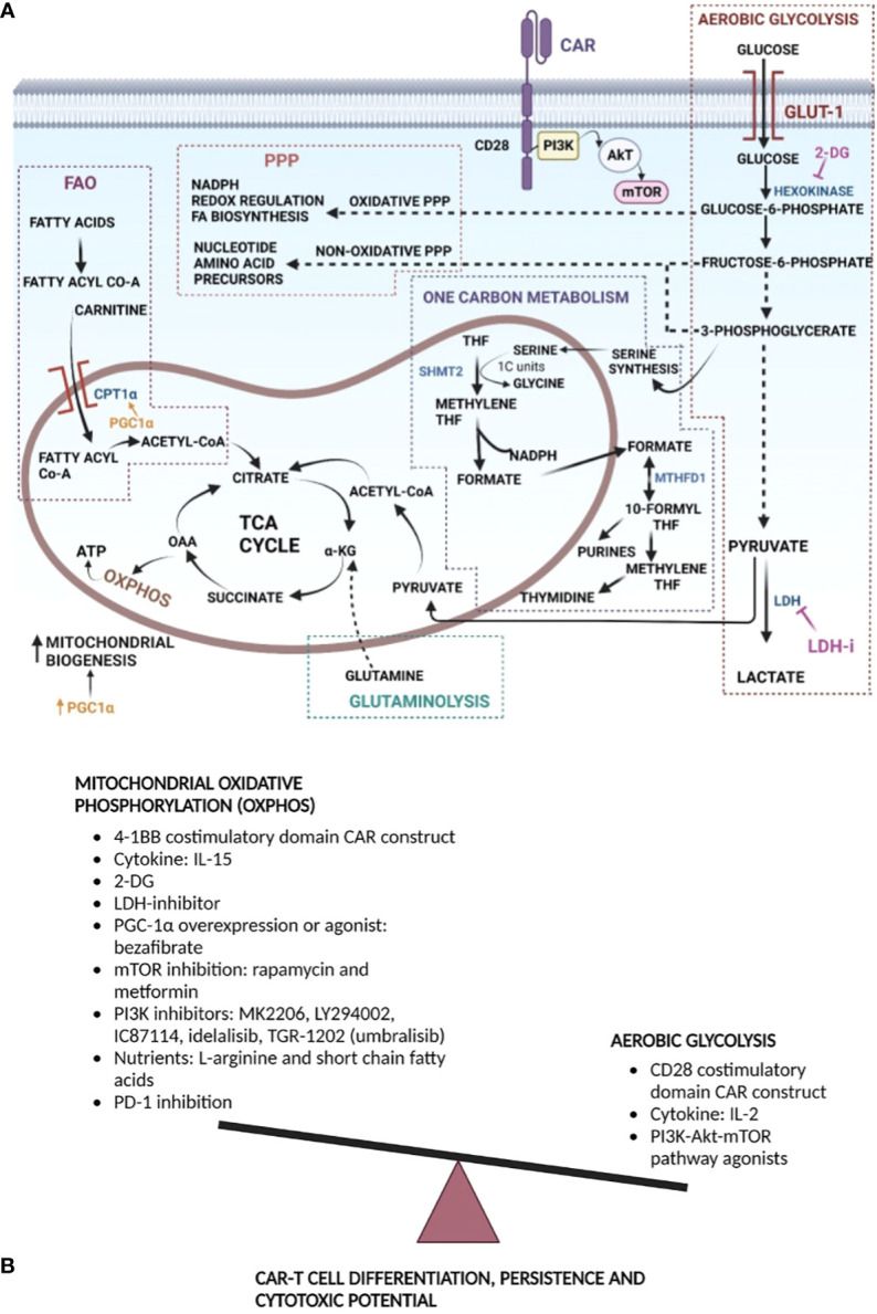
Dr. Buxbaum and her team are also exploring deuterium imaging for tracking chimeric antigen receptor (CAR) T cells in vivo and assessing their function/activation within the tumor microenvironment (TME).
CAR T cells are a form of immunotherapy, where the patient’s own T cells are engineered to kill cancer. To develop more effective CAR T therapies, tools for tracking these cells (to the tumor) after infusion and assessing their in vivo function (within the TME) are greatly needed.
Read the research
- Brender JR, et al. In vivo deuterium magnetic resonance imaging of xenografted tumors following systemic administration of deuterated water. Sci Rep. 2023 Sep 7;13(1):14699.
- Nanjireddy PM, Olejniczak SH, Buxbaum NP. Targeting of chimeric antigen receptor T cell metabolism to improve therapeutic outcomes. Front Immunol. 2023 Mar 14;14:1121565.
Connect with the Buxbaum Lab
Phone: 716-845-8151
Office Location: Cancer Cell Center 521E
Department of Pediatric Oncology
Roswell Park Comprehensive Cancer Center
Elm and Carlton Streets
Buffalo, NY 14263