If your doctor suspects a spinal tumor, you will likely undergo one or more of the following:
- Complete physical and neurologic exam, including a complete health history
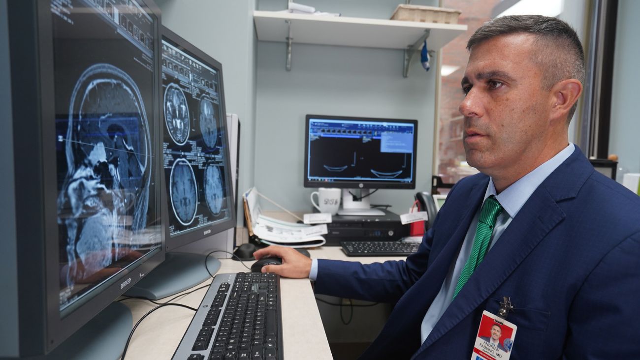
- Magnetic resonance imaging (MRI): This type of body scanning uses an SUV-size device, built around a powerful magnet and linked to a computer, to take detailed pictures of the interior of your spine. In some cases, a harmless dye called contrast is injected through a blood vessel in your arm or hand to help highlight abnormal areas of the spine.
- Computerized Tomography (CT) scan/3-D imaging: An advanced, computer-assisted X-ray machine takes detailed pictures of the spine that can be used in surgical planning.
- Nuclear medicine bone scan: A small amount of radioactive material is injected into a blood vessel, and it travels through the bloodstream to the bones. This scan can show if cancer has spread to the bone.
- Biopsy: A sample of the suspicious tissue may be taken with a small needle under X-ray guidance, or during a larger surgical procedure to correct the symptoms caused by the tumor. A biopsy is the most certain way to diagnose and plan treatment for a spinal tumor if the tumor type is unknown. However, in patients who are already known to have metastatic disease — for example, metastatic breast cancer — often there is no need to perform another biopsy. It’s presumed that the spinal tumor spread from the breast, so treatment can begin without delay.
When a spinal tumor is suspected, Roswell Park’s Neuro-Oncology team gathers as much information as possible to confirm the diagnosis and describe the tumor: How large is it? Is it malignant (cancer) or benign (not cancer)? Did it start in the spine or travel to the spine from another part of the body? Our experts in Neuroradiology and Neuropathology help answer those questions to determine your best treatment options.