How is liver cancer diagnosed?
If you experience any symptoms (such as discomfort in the right upper part of the abdomen) or abnormal liver function blood test results, your physician will likely order medical imaging to examine the liver. Some cases are found incidentally during an ultrasound, CT scan or MRI scan that was ordered for another reason.

People who face a higher risk for liver cancer due to hepatitis or cirrhosis, may have routine screening tests such as blood tests to measure alpha-fetoprotein (AFP) or an ultrasound of the liver.
It matters who performs and reads your tests
Not all liver problems are due to cancer, and many liver conditions can appear cancer-like, such as hepatic hemangioma, focal nodular hyperplasia, hepatic adenoma, nodular regenerative hyperplasia and regenerative nodules. Careful review of properly performed imaging is key to assessing tumor growth over time and appearance of blood flow and other structures. Roswell Park’s diagnostic radiologists and pathologists are highly experienced in determining whether a liver condition is malignant.
To arrive at a definitive diagnosis, you may undergo one or more of these tests:
- Ultrasound. Uses sound waves to produce pictures of internal body structures.
- Blood tests. Measuring certain substances in the blood, such as AFP, can be used to assess liver function.
- Multiphasic high-definition “liver protocol” CT. This focused scan produces high-resolution images specifically of the liver.
- Magnetic resonance imaging (MRI) and magnetic resonance cholangiopancreatography (MRCP). These MRI scans use powerful magnets and radio waves to take clear pictures of the liver, pancreas and bile ducts.
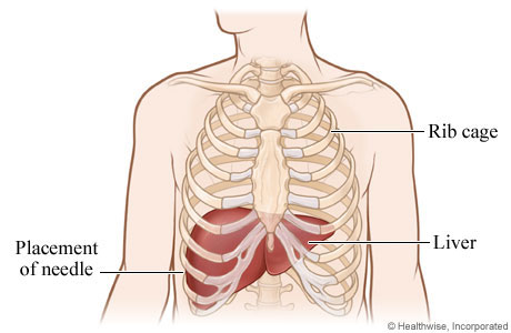
- Image-guided biopsy. This procedure uses CT and ultrasound images to obtain a small sample of the tumor to analyze and learn whether it contains any cancer cells.
- Diagnostic laparoscopy with intraoperative ultrasound. In this procedure, small incisions are made in the abdomen, and a laparoscope (a thin, lighted tube) and ultrasound devices are inserted to produce internal pictures.
- Bone scan. This type of imaging can detect whether cancer has spread to the bones.
You have time for a second opinion
A correct diagnosis of your cancer is essential to your best chance for a cure. About 10% to 18% of the outside cases we review at Roswell Park result in a change of diagnosis — either a reversal of diagnosis (someone received a cancer diagnosis but does not actually have cancer, or someone has cancer but was told no cancer was present) or our pathologists identify a type of cancer that is different from the one that was originally diagnosed; this information can dramatically alter the treatment plan. Seek a second opinion at Roswell Park before making any surgery or treatment decisions.
