Diagnostic and staging tests
Determining a diagnosis of esophageal cancer requires state-of-the-art imaging and diagnostic tests plus the expertise of diagnostic radiologists, pathologists and oncologists who specialize in upper gastrointestinal cancers. In order to reach an accurate diagnosis and determine whether and where the cancer may have spread (called staging), you may undergo one or more of the following tests or procedures:
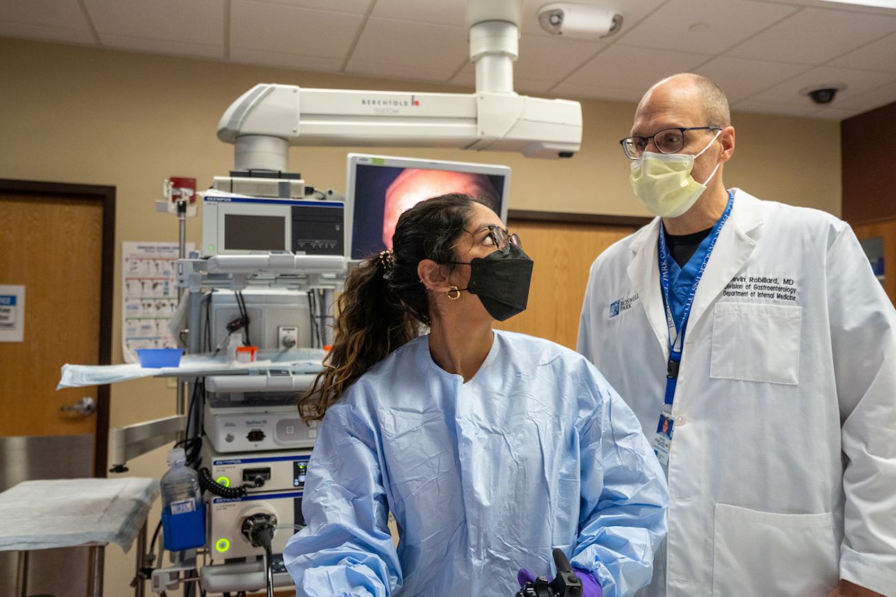
- Upper endoscopy. After numbing your throat with an anesthetic spray, a thin, lighted tube (endoscope) is passed through your mouth or nose into the esophagus so the physician can examine the inside of the esophagus.
- Barium swallow or “upper GI series.” After drinking a barium solution, x-rays are taken of your esophagus and stomach. The solution highlights the esophagus on the x-rays.
- Endoscopic ultrasound (EUS). Performed only at specialized centers, this procedure uses a tool similar to the endoscope, but with a small ultrasound device at the tip. EUS provides high-quality and detailed images useful for diagnosing and staging upper GI cancers. A needle may be used to tissue samples of lymph nodes.
- Computed Tomography (CT) scan. An x-ray machine linked to a computer takes a series of detailed pictures of your chest and abdomen to learn whether the cancer has spread to lymph nodes and other areas. You may receive contrast material by mouth or by injection into a blood vessel, to highlight any abnormal areas.
- Image-guided biopsy. Performed with the assistance of an ultrasound machine or CT scan.
- Magnetic Resonance Imaging (MRI) scan. A powerful magnet linked to a computer is used to make detailed pictures of areas inside your body to show whether cancer has spread to lymph nodes or other areas. Sometimes contrast material is given by injection into a blood vessel. The contrast material makes abnormal areas easier to see.
- Positron Emission Tomography (PET) scan. You receive an injection of a small amount of radioactive sugar. The sugar gives off signals that the PET scanner picks up. The PET scanner detects areas in your body where the sugar is being taken up. Cancer cells show up brighter in the picture because they use energy (sugar) faster than normal cells do. A PET scan shows whether esophageal cancer may have spread.
- Laparoscopy. After you are given general anesthesia, the surgeon makes small incisions (cuts) in your abdomen. The surgeon inserts a thin, lighted tube (laparoscope) into the abdomen. Lymph nodes or other tissue samples may be removed to check for cancer cells. Sometimes staging tests are not complete until after surgery to remove the cancer and nearby lymph nodes.
Request an appointment Treatment for esophageal cancer
Tumor biomarker testing
Specialized physicians, called pathologists, will examine and test the tissue or tumor that was removed by biopsy or surgery to determine whether it has certain biomarkers. The result of these analyses can help determine whether new treatments that target these biomarkers can help treat your cancer. These markers include:
- HER2 (Human epidermal growth factor receptor 2). Some esophageal tumors have too much of this protein which causes them to grow and spread quickly. A drug called trastuzumab targets this protein on the cancer cells.
- PD-L1 (programmed death ligand-1) is a protein sometimes found in cancer cells that prevent your body’s immune system from recognizing and attacking the cancer. Immunotherapy drugs that interfere with PD-L1, such as checkpoint inhibitors, may help your immune system fight your cancer.
- Microsatellite instability or mismatch repair. This marker points to a genetic defect that means your genes are unable to repair damaged DNA. If testing shows you have high microsatellite instability or are mismatch repair deficient, immunotherapy with checkpoint inhibitors may be part of your treatment plan.