Anatomic and functional imaging of prostate tumors
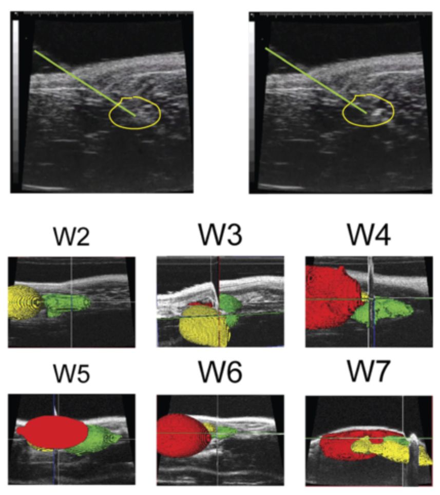
The Nastiuk Lab has developed high-resolution anatomic imaging to better understand how prostate tumors respond. We refined both HFUS and MR anatomic imaging to enable accurate and precise reproducible serial tumor volume determination.
We also employ three functional imaging techniques:
- Power Doppler to assess blood flow
- Photoacoustic imaging to assess tumor O2 saturation
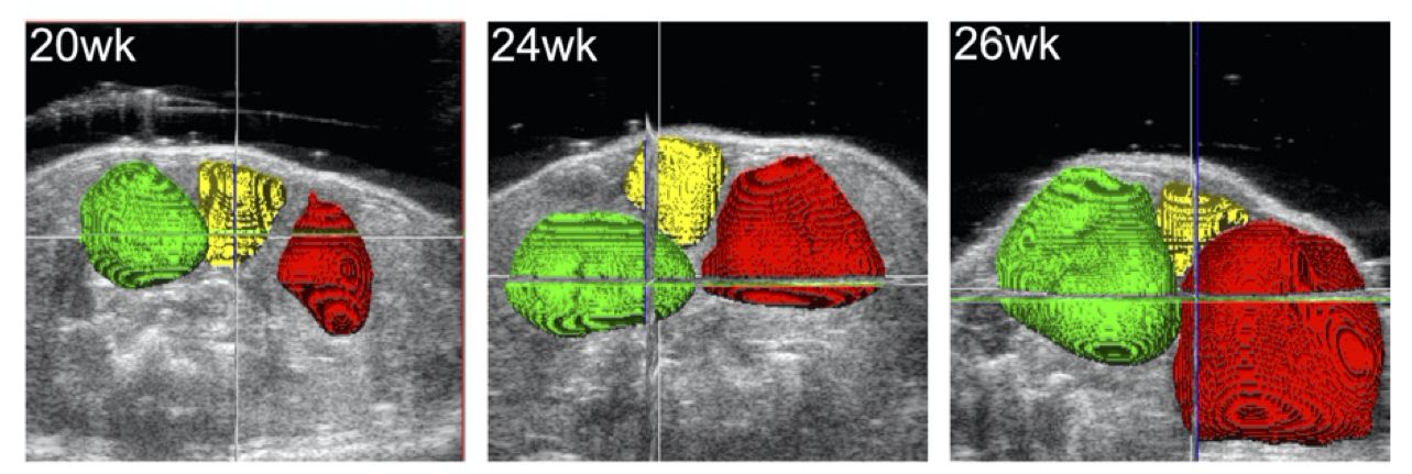
- Contrast-enhanced ultrasound to determine tissue perfusion
Using these imaging modalities, we can measure both anatomic (tumor volume) and functional responses (e.g., hypoxia), to experimental manipulations (e.g., novel inhibitors), that may inform therapy.
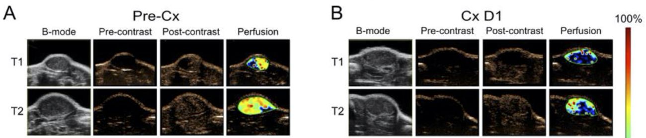
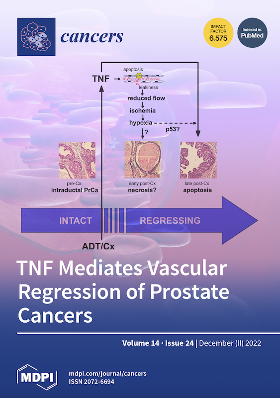
TNF mediates vascular regression of prostate tumors
Androgen deprivation therapy (ADT) is the principal therapy for advanced prostate cancer. ADT controls tumor growth by rapidly altering the prostate tumor microenvironment and subsequently inducing cancer cell death. ADT induces vascular damage and thereby reduces intratumoral blood flow, but the mechanism has long been elusive.
Employing our anatomic and functional imaging we showed TNF acts as the mediator of castration-induced vascular damage in prostate tumors. This pathological response to androgen deprivation—beginning with endothelial cell apoptosis and increased vessel permeability and culminating in hypoxia indirectly contributes to prostate cancer regression.
Because TNF is also a critical death receptor ligand for prostate epithelial cells, we propose that TNF is a multi-purpose, comprehensive signal that mediates prostate cancer regression following androgen deprivation.
See the science
- Krolewski JJ, Singh S, Sha K, Jaiswal N, Turowski SG, Pan C, Rich LJ, Seshadri M, Nastiuk KL. TNF Signaling Is Required for Castration-Induced Vascular Damage Preceding Prostate Cancer Regression. Cancers (Basel). 2022 Dec 7;14(24):6020. doi: 10.3390/cancers14246020.
- Schmitthenner HF, Dobson DE, Jones KG, Akporji N, Soika DQM, Nastiuk KL, Hornak JP. Modular Synthesis of DOTA-Metal-Based PSMA-Targeted Imaging Agents for MRI and PET of Prostate Cancer. Chemistry. 2019 Nov 4;25(61):13848-13854. doi: 10.1002/chem.201903390.
- Dogra V, Chinni B, Singh S, Schmitthenner H, Rao N, Krolewski JJ, Nastiuk KL. Photoacoustic imaging with an acoustic lens detects prostate cancer cells labeled with PSMA-targeting near-infrared dye-conjugates. J Biomed Opt. 2016 Jun 1;21(6):66019. doi: 10.1117/1.JBO.21.6.066019.
- Singh S, Pan C, Wood R, Yeh CR, Yeh S, Sha K, Krolewski JJ, Nastiuk KL. Quantitative volumetric imaging of normal, neoplastic and hyperplastic mouse prostate using ultrasound. BMC Urol. 2015 Sep 21;15:97. doi: 10.1186/s12894-015-0091-9.
Connect with the Nastiuk Lab
Department of Cancer Genetics & Genomics
Roswell Park Comprehensive Cancer Center
Elm and Carlton Streets
Buffalo, NY 14263