Get yourMammogramwhere the experts are.
Call 1-800-767-9355
No prescription or referral needed to make an appointment
Did you know that annual screening mammography saves lives?
- 1 in 8 women will get breast cancer in their lifetime
- Your best fighting chance against breast cancer is regular screening mammograms starting at age 40
- Most mammograms are normal… but if yours isn’t, Roswell Park is where you want to be
- Our team of experts deal only in breast imaging, allowing them to identify and determine results confidently
No prescription or referral needed to make an appointment
Even if you have a prescription for another location for your mammogram, or have gone elsewhere previously, we are able to get your scans for comparison.
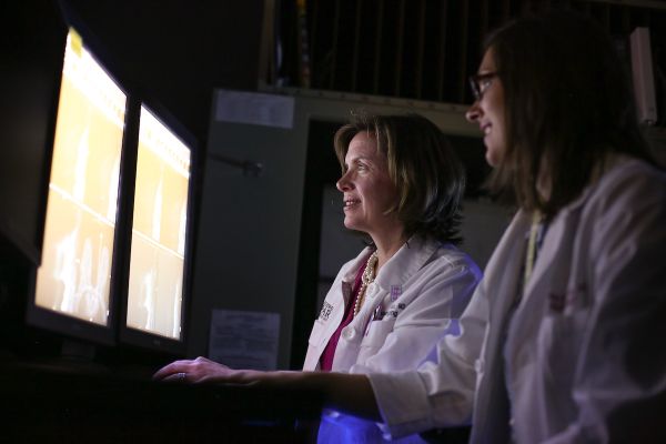
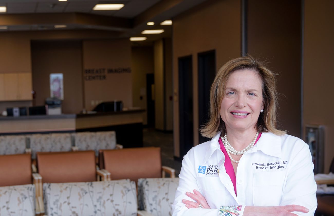
Experts in cancer imaging
It matters where you get your mammogram and it matters who reads your mammogram. We have a dedicated team of technologists and radiologists who specialize in the detection of breast cancer. Most mammograms are normal, but if yours isn’t, this is where you want to be.
3D mammography
Our Scott Bieler Clinical Sciences Center houses a spacious state-of-the-art Breast Imaging Center equipped with digital mammography and tomosynthesis (also known as 3D screening) capabilities.
Locations & Hours
Downtown
Appointments for screening mammograms are available Monday - Friday, between 7:30 a.m. and 4 p.m.
Later appointments can be made available upon request.
955 Michigan Ave,
Buffalo, NY 14203
Free, convenient parking is provided for screening mammograms in the lot at the corner of Michigan Street and Carlton Street (look for the pink sign).
Breast Care of Western New York
Appointments for screening mammograms are available Monday - Friday, between 7:45 a.m. and 3 p.m.
199 Park Club Lane, Suite 100,
Williamsville, NY 14221
Community Outreach Programs
✔ Esperanza y Vida – offering translation, patient navigation, and screening assistance to our Hispanic/Latinx community. Call 716-845-4623.
✔ National Witness Project – educates participants in early cancer detection in churches and community settings, offering patient navigation and screening assistance. Call 716-845-1394.
Have a family history of breast cancer?
Learn about our high-risk program.
The Breast Cancer Risk Assessment & Prevention Program offers the most advanced surveillance, screening, diagnostic and preventive methods to women most at risk for breast cancer.
To learn more about the program, or whether you qualify, call 1-800-ROSWELL (1-800-767-9355) or complete an online form.