Roswell Park recently became the first cancer center in Western and Upstate New York to use a technology that allows neurosurgeons enhanced views of brain tumors during surgery.
The new surgical visualization system provides magnification with greater detail and a wider view of the operative field than ever was possible before.
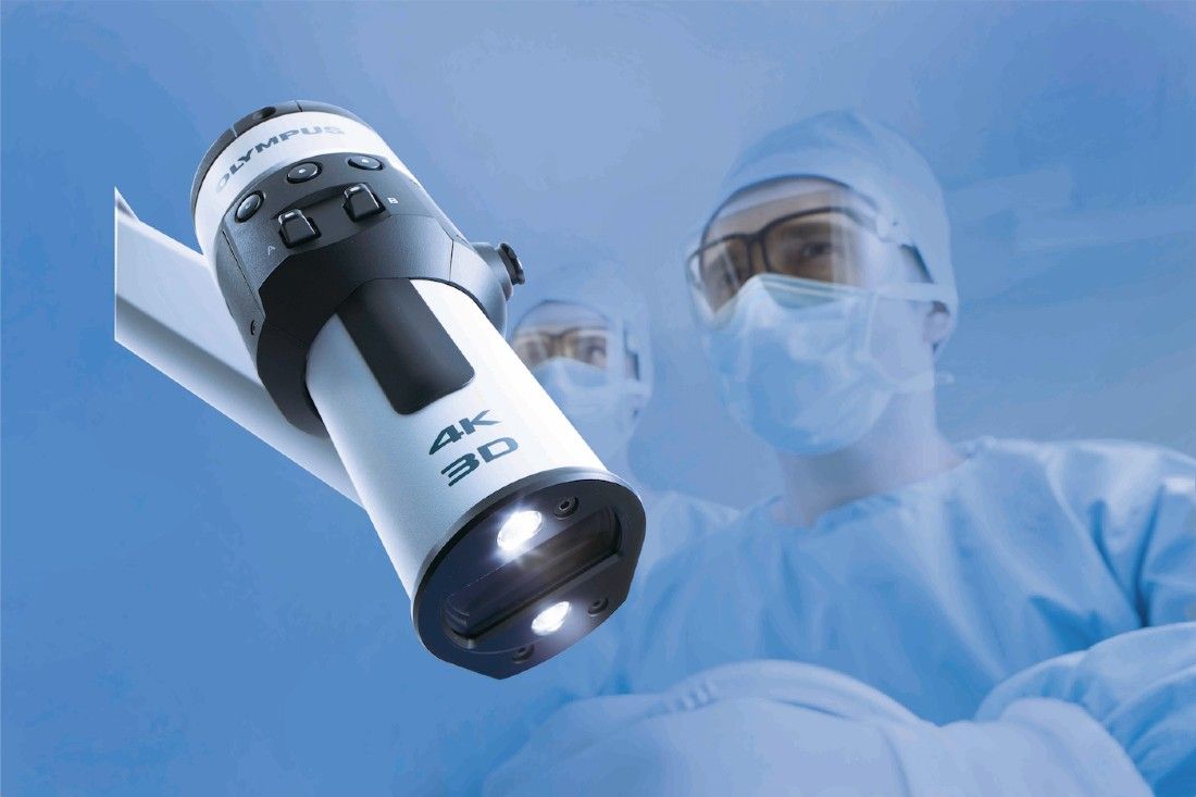
The system is called the Olympus ORBEYE®. A monitor in the operating room combines two cameras with advanced image processing to give doctors a 3D TV-screen view of micro-neurosurgical anatomy, the very fine details of the brain that are magnified during surgery.
Neurosurgeons wear high-quality 3D glasses to view the screen. This creates a big-screen effect through 4K and 3D advanced technology that improves visualization as they work. Higher resolution and a broader color spectrum help them to better identify the boundaries between tissue, blood vessels and lesions. Being able to see the transition between tumor and normal brain is of particular importance in these surgeries, because it allows maximal removal of the tumor without damaging healthy tissue nearby.
“It does take some getting used to, as the surgeon is not looking directly in the wound but at a TV screen while performing the operation,” notes Robert Fenstermaker, MD, Chair of Roswell Park’s Department of Neurosurgery. “It means retraining your brain to learn hand-eye coordination.”
Dr. Fenstermaker says the technology has transformed the way he and other neurosurgeons perform surgeries and help their patients. “Enhanced visualization technology provides an unparalleled view of tumors and normal brain anatomy during surgery,” he says. “Previous visualization methods were bulky, limiting freedom of movement in the surgical field. Now the surgeon can ‘look around corners’ during surgery in an unprecedented way.”