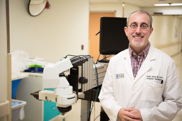
Dr. Skitzki's microscopy system is the first of its kind
A brand new innovation developed at Roswell Park Comprehensive Cancer Center may change the way we look at cancer tumors — literally.
In addition to treating melanoma and sarcoma patients at Roswell Park, Joseph Skitzki, MD, FACS, spent the last few years developing a high-powered, first-of-its-kind microscope for use in the operating room. In February 2016, following a short study of the microscope’s functionality, Dr. Skitzki's research team revealed its stunning findings.
The objective behind creating the microscope was to get a clearer glimpse inside the human body. Most microscopes used during surgical procedures magnify up to 15x – Dr. Skitzki’s high-power intravital microscopy (IVM) system operates at 100x magnification or higher, providing an unprecedented view of tumor blood vessels.
IVM, previously only used in animals, provides an up close view of human tumors in real time, offering new insights into their size and how they function. Tumors are currently viewed only in pathology following physical removal via surgery.
"We are excited about the possibilities," said Dr. Skitzki. "Our next clinical trial will determine our ability to look at lymph nodes in melanoma patients as they are often the first site of tumor spread."
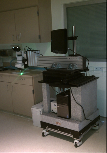
When picturing a microscope, your mind most likely forms an image of the traditional version found in laboratories and classrooms across the world.
The microscope used to perform IVM has a slightly different appearance.
Stabilized by a mobile 800-lb. granite platform, Dr. Skitzki’s invention utilizes digital video and an epifluorescent light source to magnify its subjects. During surgery, a mechanical arm attached to the microscope extends over the patient, recording live, high-definition footage of cancerous tumors.
"There were lots of different design proposals," said Dr. Skitzki. "Initially we thought maybe there was something we could hook up to the operating room table, but that would not be stable enough. Over the course of a year, we settled on this design."
The initial study performed by Dr. Skitzki and his team revealed three important takeaways:
- IVM is safe and feasible to perform on humans
- Approximately half of all tumor blood vessels are non-functional
- Human tumor blood vessels are wider than previously thought via pathological study
Blood vessels are the major route for almost all cancer treatments. If only half of the vessels are open, parts of the tumor exist that are never exposed to these treatments. Additionally, the finding of wider blood vessels may influence calculations regarding delivery of drugs and immune cells to the tumor, which was previously unknown.
The researchers will continue to examine the microscope’s efficacy through clinical studies, with the hope that applying IVM to surgery on humans will revolutionize cancer treatment while also improving outcomes.
"In the future, we hope to develop reagents to use in combination with the microscope to better define responses to treatments and tailor therapies for each individual cancer patient," said Dr. Skitzki. "Right now, however, we are still in the clinical trials phase of development.”
Dr. Skitzki's innovation was made possible in part by the Jennifer Linscott Tietgen Family Foundation, whose generous donation paid for the microscope in full. The Tietgens began the Foundation in memory of their daughter, Jennifer, and are dedicated to supporting melanoma cancer research.
“We knew we needed to do something to make a difference, and the best way we could think of was funding melanoma research projects," the Tietgens told Roswell Park in February 2016. "The Roswell Park melanoma team has an advanced understanding of this devastating cancer and is working tirelessly to improve diagnostic testing and treatment.”
The work was also supported in part by three National Institutes of Health grants from the National Institute of Allergy and Infectious Diseases (NIAID).