How is pancreatic cancer diagnosed?
If you have a suspicion of pancreatic cancer, you need a prompt and accurate diagnosis. Roswell Park’s team of experienced diagnostic radiologists and pathologists who focus on pancreatic cancer can provide the precise answers you and your physicians need to determine your treatment options. To arrive at an accurate diagnosis, you may undergo one or more of the following:
- Pancreas protocol computed tomography (CT) scan is a CT scan that focuses on the pancreas. CT uses an x-ray machine linked to a computer to take a series of high-resolution pictures. The computer puts the x-rays together to create images of the pancreas and other organs and blood vessels in the abdomen.
- Pancreas protocol magnetic resonance imaging (MRI) uses a powerful magnetic field and radio waves to produce clear, detailed images of the pancreas. MRI does not use x-rays.
- Magnetic Resonance cholangiopancreatography (MRCP) is a magnetic resonance imaging (MRI) scan that uses powerful magnets and radio waves to take clear pictures of the pancreas and bile ducts. No contrast dye or radiation is needed with MRCP.
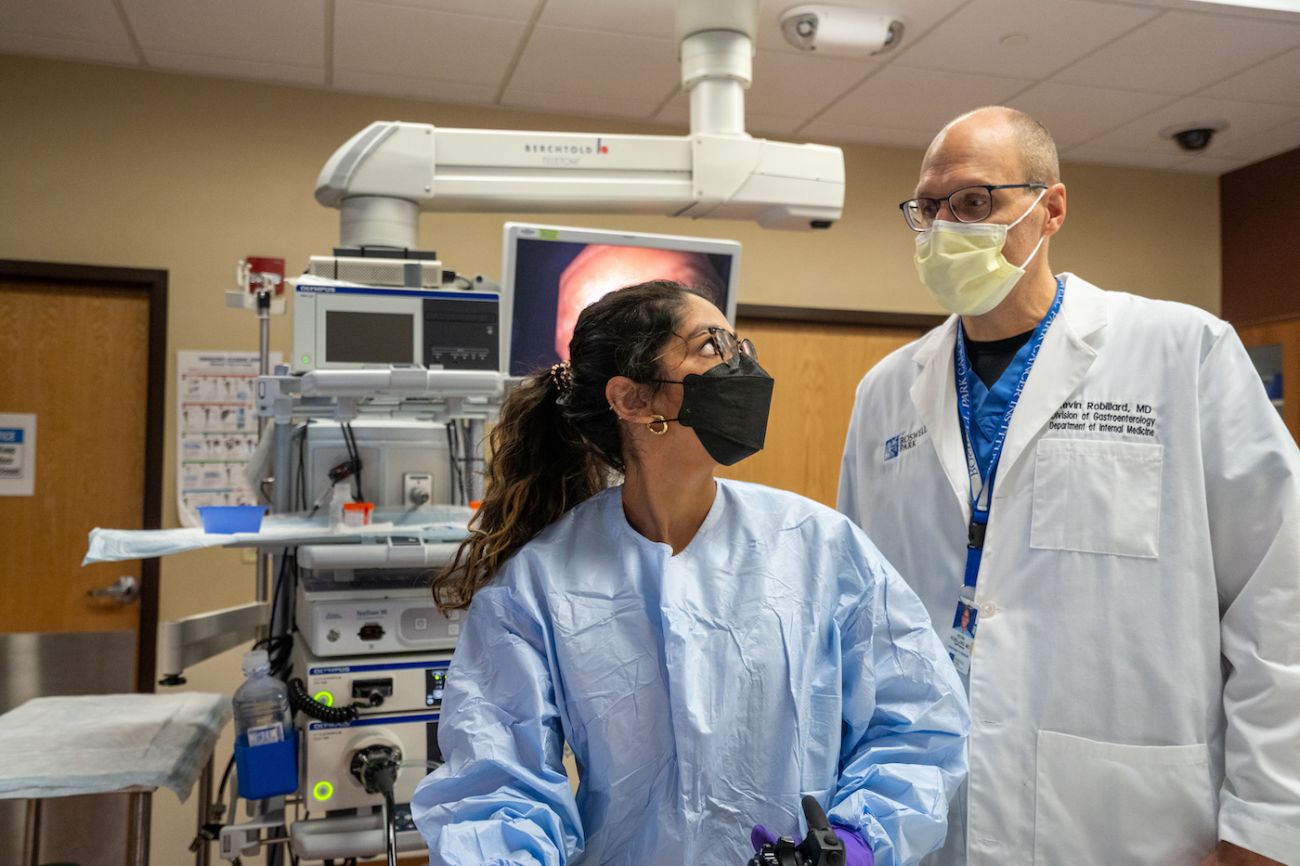
- Endoscopic ultrasound (EUS) uses a thin, lighted tube (endoscope) passed through the patient's mouth and stomach into the first part of the small intestine. At the tip of the endoscope is an ultrasound device. The doctor slowly withdraws the endoscope from the intestine toward the stomach to make images of the pancreas and surrounding organs and tissues. A biopsy may be taken during this procedure to confirm a diagnosis.
- Endoscopic retrograde cholangiopancreatography (ERCP) uses an endoscope passed through the patient's mouth, stomach, and into the first part of the small intestine. Then, a smaller tube (a stent) is passed through the endoscope and into the bile ducts and pancreatic ducts. Dye is injected through the catheter to the duct, and x-rays can show whether the ducts are blocked from a tumor or other condition.
- Percutaneous transhepatic cholangiography (PTC) is a procedure that involves injecting a dye through a thin needle inserted through the skin into the liver. On x-ray, the physician can see whether the dye moves freely through the bile ducts, or whether they are blocked from a tumor or other condition.
- Image-guided biopsy obtains a small piece of the tumor under the guidance of imaging scans, such as CT, MRI and ultrasound. As a high-volume center, our experienced physicians perform this accurately and expediently, allowing faster treatment planning.
- Blood tests to measure the number of specific cells or the level of certain chemicals or other substances can indicate whether pancreas cancer may be present or assess your response to treatment. These may include:
- CA 19-9, a blood protein that may be elevated in people with pancreas cancer.
- Bilirubin, a substance in bile that may reach high levels in the blood if the bile duct is blocked. High bilirubin in the blood causes jaundice and increased CA 19-9 levels.
- Complete blood count will reveal whether you have too few or too many of certain blood cells, which may suggest cancer and other conditions.
What comes next after diagnosis?
The Endoscopy Center
Our Endoscopy Center brings together Advanced Endoscopy and Interventional Pulmonology services into one facility where more than 90% of procedures, including pancreatic cancer screening exams, are performed on an outpatient basis.