In determining whether a mole or skin lesion contains any malignant cells, doctors take a biopsy — removing all or part of the abnormal area — and send to a pathologist for examination and testing.
For suspected melanomas, your physician will likely do one of these procedures:
- Shave biopsy – a thin, sharp blade shaves off the abnormal tissue.
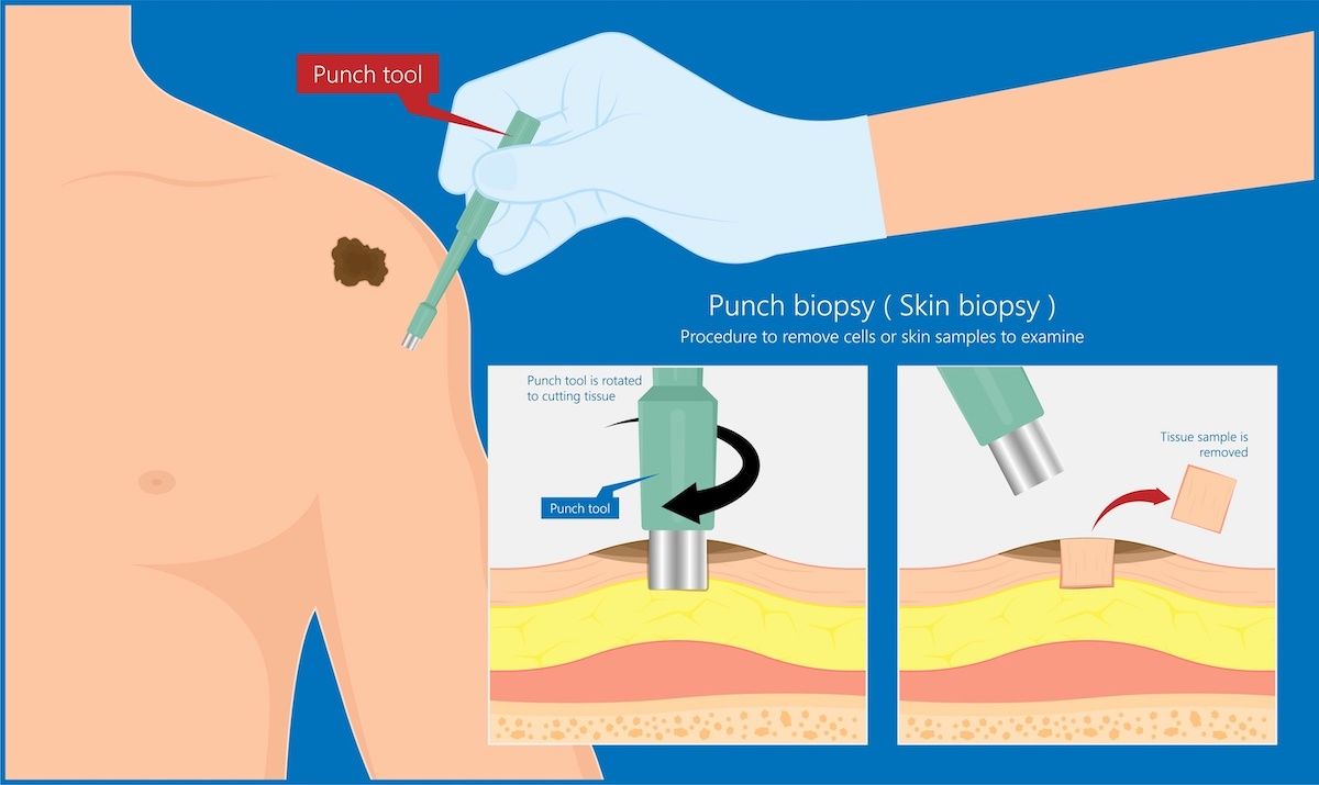
- Punch biopsy – a sharp, hollow device removes a small but deep sample of tissue.
- Excisional biopsy – The physician uses a scalpel to remove the entire mole or growth and some tissue around it. You will have local anesthesia, so you won’t feel pain.
- Incisional biopsy – This type of biopsy where only a piece of the lesion or mole is removed may be done if the area of concern is very large or difficult to remove completely.
Understanding your pathology report
In analyzing your biopsy, these factors also play a role in diagnosing and staging your cancer:
- Breslow thickness – How deep (in millimeters) the tumor has grown into the layers of the skin.
- Ulceration status – whether or not the tumor’s top skin layer has eroded.
- Dermal mitotic rate – the number of growing tumor cells found in the tissue sample.
- Margin status – whether any cancer cells remain in the tissue surrounding the sides of the tumor (peripheral margin) or in the tissue under the tumor (deep margin).
- Microsatellites – This refers to the presence (or absence) of tiny tumors are found near the main tumor, but are separate (actually lymphatic spread).
- Pure desmoplasia – whether or not the melanoma is a rare type called desmoplastic melanoma, which may be further classified as pure or mixed.
- Lymphovascular or Angiolymphatic invasion – whether the cancer has invaded lymph nodes or blood vessels.
Other diagnostic tests
Because melanoma is more likely to spread than other skin cancers, you may have additional tests and imaging to learn whether the cancer has grown deeper in the skin, spread to lymph nodes, or other parts of the body such as the lungs, or shows specific characteristics or mutations, The information learned from these tests helps guide which treatment options will work best against your melanoma. These additional tests may include:
- Computerized tomography (CT)
- Magnetic resonance imaging (MRI)
- Positron Emission tomography (PET)
- Immunohistochemistry staining to determine if the melanoma cells have a specific characteristic such as BRAF mutation.
- Lymph node testing
- Sentinel lymph node biopsy – removing cells, a portion of the node or the entire lymph node most likely (such as closest in proximity) to contain cancer
- Fine needle aspiration – uses a thin needle to remove cells from a lymph node
- Excisional lymph node biopsy – removes a portion of a lymph node