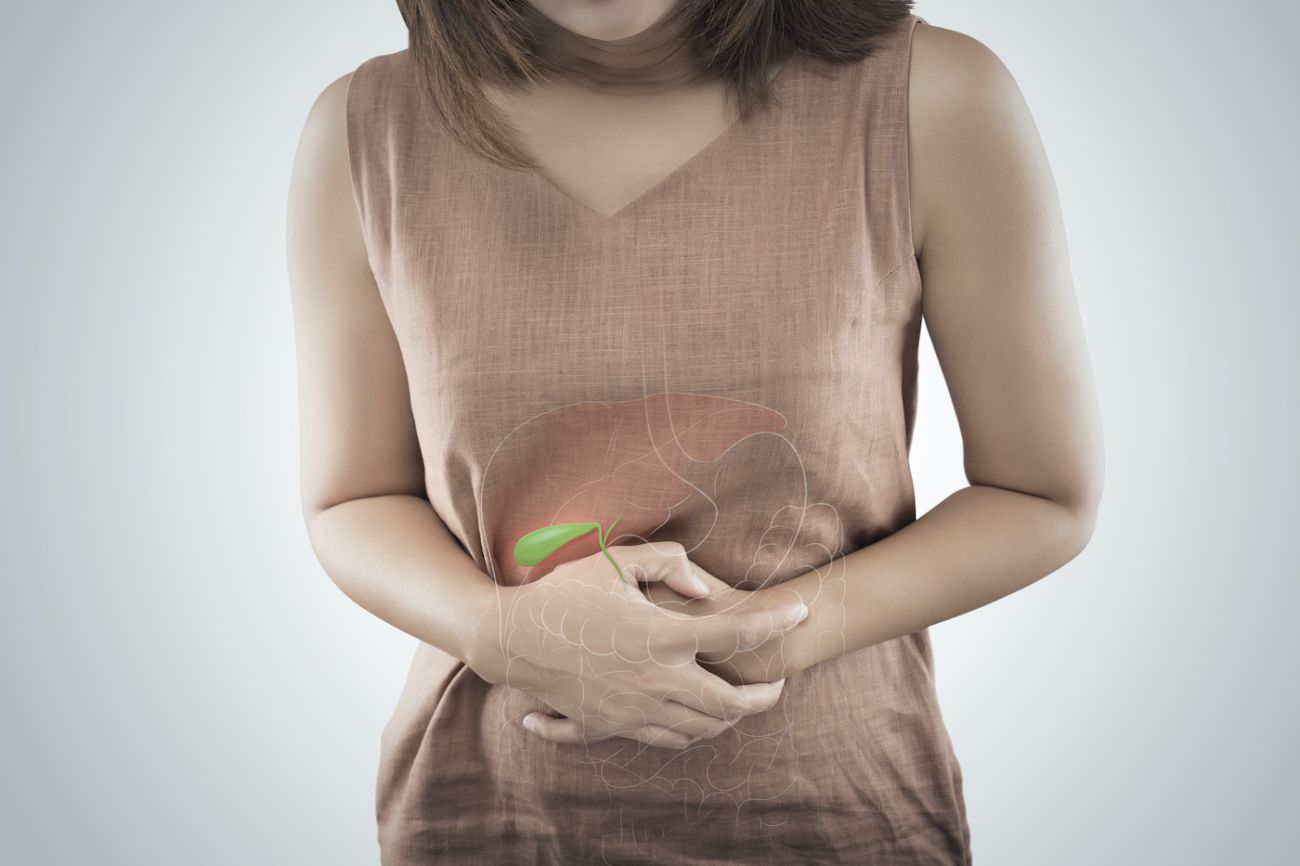
If you have a suspicious mass in your gallbladder or bile duct, it’s important to be evaluated by a cancer expert before you make any further decisions. A biopsy may be unnecessary and may risk dissemination of cancer cells.
Gallbladder and bile duct cancers are difficult to detect at an early stage for several reasons: symptoms are often vague and mimic other illnesses; the gallbladder and bile ducts are hidden beneath the liver; and screening tests to detect cancer before symptoms appear are not yet developed. In many cases, these cancers are found incidentally, as a result of imaging scans or other tests done for another reason, or surgery to remove gallstones.
Occasionally the diagnosis is unclear from imaging and laboratory tests and a biopsy is still needed, which can be performed safely by our skilled interventional radiologists. To confirm your diagnosis, assess disease stage and plan appropriate treatment, you will likely have one or more of the following:
Liver function tests
Higher than normal blood levels of substances made by the liver can indicate liver problems that may be caused by gallbladder or bile duct cancer.
Tumor marker tests
The amounts of certain substances are measured in samples of blood, urine or tissue. Elevated levels of carcinoembryonic antigen and CA 19-9 are associated with gallbladder and bile duct cancers.
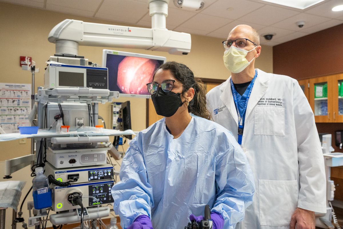
Endoscopic Retrograde Cholangiopancreatography (ERCP)
This x-ray of the bile ducts can reveal whether gallbladder or bile duct cancer has narrowed or blocked the ducts. A thin, lighted tube (endoscope) is passed through the mouth, esophagus, and stomach into the first part of the small intestine. A smaller tube (catheter) inserted through the endoscope into the bile ducts delivers dye to the ducts and an x-ray is taken. If ducts are blocked, a fine tube may be inserted to unblock the duct, or placed (as a stent) to keep the duct open. Tissue biopsy may be done during this procedure.
Endoscopic Ultrasound (EUS)
A thin, lighted tube (endoscope) is passed through the patient's mouth and stomach into the first part of the small intestine. At the tip of the endoscope is an ultrasound device. The doctor slowly withdraws the endoscope from the intestine toward the stomach to make images of the gallbladder and bile duct and surrounding organs and tissues. A biopsy may be taken during this procedure to confirm diagnosis.
Percutaneous Transhepatic Cholangiography (PTC)
A thin needle inserted through the skin below the ribs injects dye into the liver, and an x-ray is taken. If a blockage is found, a stent in the liver can drain bile into the small intestine or a collection bag outside the body.
Ultrasound exam
High-energy sound waves (ultrasound) are bounced off internal tissues or organs and make echoes. The echoes form a picture of body tissues called a sonogram.
Angiographic Computed Tomography (CT) Scan
A computer linked to an x-ray machine makes a series of very detailed high resolution pictures of areas inside the body, taken from different angles. A dye may be injected into a vein or swallowed to help the organs or tissues show up more clearly.
Magnetic Resonance Imaging (MRI)
A powerful magnet, radio waves, and a computer combine to make detailed pictures of areas inside the body. This procedure is also called nuclear magnetic resonance imaging (NMRI).
Magnetic Resonance Cholangiopancreatography (MRCP)
MRI imaging where a dye is injected to in the gallbladder area to allow the gallbladder and bile ducts to be better detailed in the image. To create detailed pictures of blood vessels near the gallbladder, the dye may be injected into a vein, called magnetic resonance angiography (MRA).
Fine Needle Aspiration (FNA) biopsy
A thin needle inserted into the gallbladder, duct or lymph node during an x-ray or ultrasound to remove a sample of cells or tissue for examination under a microscope to check for signs of cancer. Biopsy may be done during PTC or ERCP.
Laparoscopy
Small incisions are made in the abdominal wall and a thin, lighted tube (laparoscope) is inserted to allow the physician to view the gallbladder and ducts, assess nearby tissues, take biopsies or remove tissue or organs.