Ask your doctor if you can have your scans performed closer to home.
Our new Scott Bieler Amherst Center offers a full range of diagnostic and interventional radiology services with state-of-the-art technology and Roswell Park expertise.
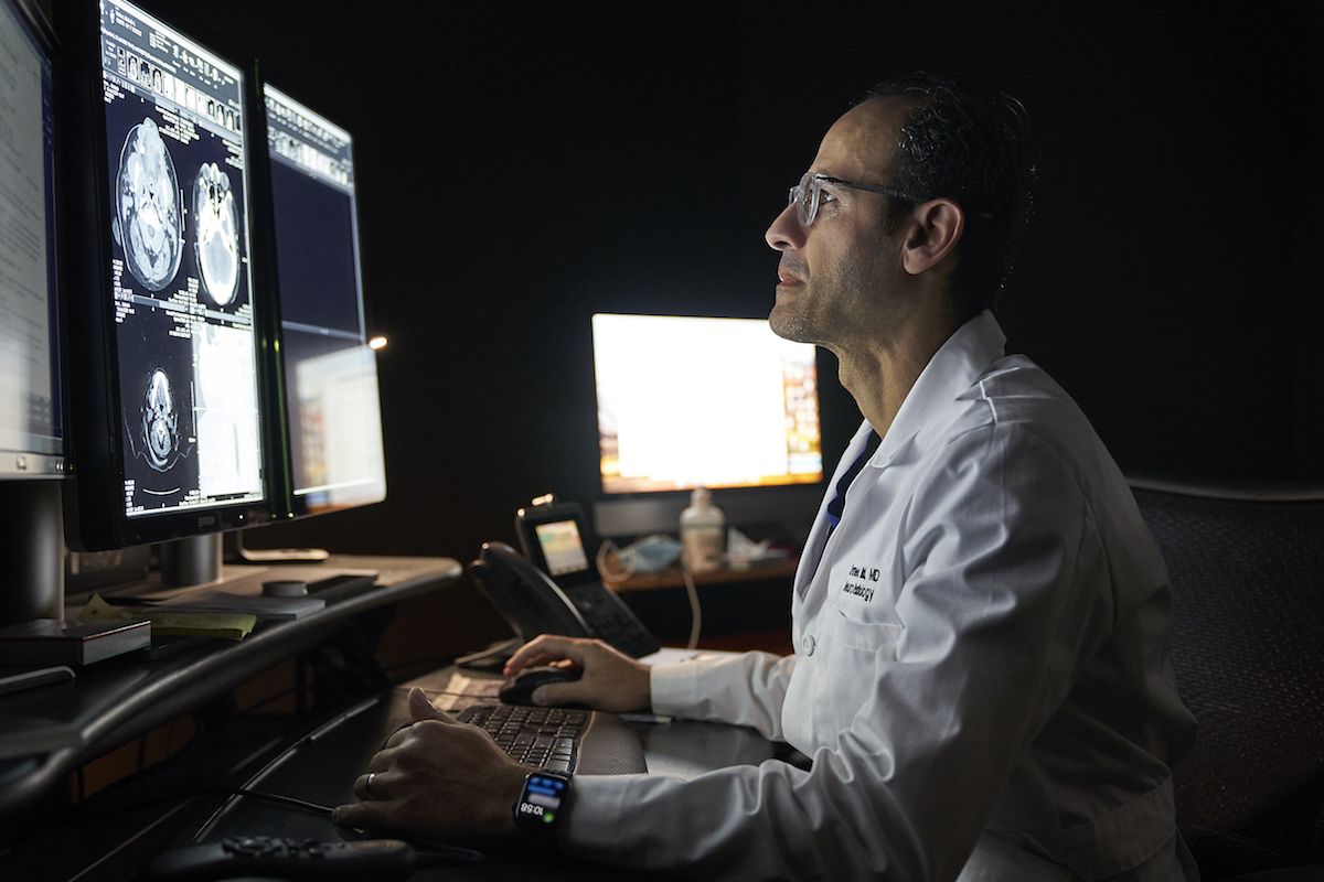
Medical imaging is a critical part of cancer care. At Roswell Park, our Department of Diagnostic & Interventional Radiology includes a team of expert radiologists uniquely experienced in medical imaging for cancer. We use the the latest technologies to diagnose cancer, guide biopsies, plan surgery and other therapies, monitor response to treatment and surveil for recurrence.
At Roswell Park, our radiologists work hand in hand with your cancer care team to provide critical information about your diagnosis and collaborate in determining your best treatment options.
Our services include
The Breast Imaging department at Roswell Park manages all breast imaging needs and related services, including:
- Screening mammography
- Breast ultrasound
- Breast MRI
- Diagnostic evaluation
- Image guided breast biopsy
- Mammography, screening and diagnostic
- Mammography 2nd Opinions and Consultations
This type of imaging scan uses an X-ray beam that rotates around the body and a computer to create detailed, cross-sectional pictures of inside the body. These cross-sectional images may be combined to create a 3D image. CT scans are fast, painless, and non-invasive. They provide detailed and accurate images with minimal ionizing radiation. CT scans are used determine whether a cancer is present, guide treatment planning and monitor treatment response. CT scans are especially useful for internal images of these body areas:
- Chest
- Abdomen
- Pelvis
- Bowel
- Head, neck and spine
- Extremities
The Diagnostic Radiology department performs the critical scans and provides diagnostic expertise to detect early signs of cancer before a person experiences symptoms. These images are especially important for people at increased risk for cancer and need enhanced surveillance. Roswell Park has dedicated programs for patients at high risk for these cancers:
Interventional radiology takes imaging a step further, using advanced medical imaging such as ultrasound and computed tomography (CT) to intervene and perform procedures such as taking a biopsy, inserting stents or chest ports, or delivering a therapy directly to the tumor.
This type of imaging uses a powerful magnetic field and radio waves to produce detailed cross-sectional images inside the body and head. These images help determine whether a tumor is present and guide future biopsies.
MRI scans are non-invasive, and unlike X-rays or CT scans, do not use ionizing radiation. Some MRI exams require a contrast solution that you may either drink or have delivered through an intravenous (IV) line in your arm. The duration of the exam varies but the average is about 30-60 minutes.
MRI’s are especially useful in getting images of the full body as well as specific body areas such as the arms, legs, heart, brain, spine, breasts, liver, biliary tract, prostate, and rectum.
Magnetic resonance cholangiopancreatography (MRCP) is a specialized type of MRI that focuses on the digestive system and creates detailed images of the liver, bile ducts, gallbladder and pancreas.
Neuroradiology is a subspecialty of radiology that focuses on medical imaging for the brain, spine, head and neck. Neuroradiologists are expert physicians who help to diagnose abnormalities and disease and guide treatment and care. We provide these services:
- Brain tumor imaging. We perform MRI scans and CT scans to detect, grade, and stage brain tumors including glioma, astrocytoma, meningioma, and metastatic brain tumors. We also perform functional and perfusion MRI, MR spectroscopy, and diffusion tensor imaging.
- Brain mapping. We use diffusion tensor imaging (DTI) and functional MRI (fMRI) to create brain maps of eloquent cortex and important white matter pathways to assist neurosurgeons with surgical planning.
- Head and neck cancer imaging. CT scans are often used for initial assessment of the primary tumor, and MRI delineate tumor extent and assess for spread to soft tissues and bone marrow. PET-CT is used to evaluate for distant metastasis and recurrent disease.
- 4D CT is a specialized imaging technique that adds a fourth dimension — time — to a three-dimensional CT scan, capturing images at different points in time to see movement and changes. At Roswell Park we use 4D CT to locate abnormal parathyroid glands, aiding in the diagnosis and treatment of hyperparathyroidism.
- Image guided procedures include CT guided biopsies of spine, head and neck tumors and skull base lesions; fluoroscopic guided diagnostic spinal taps; intrathecal chemotherapy; myelograms, cisternograms and spine injections.
Nuclear medicine is a subspecialty that uses radioactive drugs (radiopharmaceuticals) in medical imaging as well as treatment procedures. Nuclear scans make pictures based on the body’s chemistry rather than the physical shapes and forms.
When a patient is given the radiopharmaceutical, or tracer, is given to the patient, some body tissues, like those affected by cancer, take up more or less of the radioactive drug than normal tissues. A special camera that detects the radioactivity then creates an image based on where the drug travels and collects in the body.
Nuclear medicine services include the following:
- PET CT
- Bone scans
- Radioiodine scans
- Radioactive iodine therapy
- Theranostic treatments
Theranostics
Theranostics is an emerging field of cancer care that uses the methods and tools of nuclear medicine to assess your cancer — and treat it with radiopharmaceuticals. Roswell Park’s new Theranostics Center expands our ability to accommodate more patients with these novel therapies.
Also known as sonography, ultrasound produces pictures of the inside of the body using high frequency sound waves instead of radiation. Some common ultrasound scans your doctor may order include:
- Abdominal and Pelvic to evaluate your liver, pancreas, gallbladder, kidneys, bladder, and uterus
- Thyroid scan to evaluate nodules or other thyroid disease
- Vascular evaluation with Doppler imaging to check for blood clots
X-rays use electromagnetic waves to create images of the body, especially useful for images of the bones, lungs and some solid organs. Fluoroscopy is a type of imaging that uses x-rays to create real-time moving images. At Roswell Park, x-ray and fluoroscopy are used for the following serives:
- Gastrointestinal evaluation
- Speech pathology swallow evaluation
- Bone density scan (DEXA)
- Routine radiographic examinations