Microscopic features of human tumors have only ever been measured with simple snapshots in time. Tumors are usually either biopsied or removed and pathologic evaluation includes fixing the specimen and then performing either stains or other types of analysis that cannot measure any dynamic features.
Intravital microscopy is a tool used in preclinical animal experiments to microscopically observe tissue in real time. Intravital microscopy has only been performed in preclinical animal experiments. Tumors grown in animals may be observed using intravital microscopy to determine such things as blood vessel characteristics, blood flow features, tumor structure, immune cell and tumor interactions, and drug delivery. These observations are performed of dynamic processes which are then video recorded and electronically stored for further analysis. The use of intravital microscopy has offered researchers a method of measuring dynamic interactions that are not possible with simple snapshots over time.
There is no current equipment or study that has been published or presented regarding human intravital microscopy.
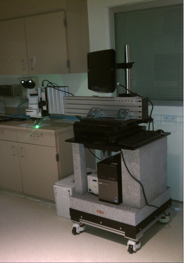
Researchers at Roswell Park Comprehensive Cancer Center have created the first intravital microscope for human use during surgical removal of tumors. The initial clinical trial has confirmed that the designed intravital microscope is fully functional and can obtain dynamic data that has never been quantified previously.
The microscope (see figure below) is constructed into a cantilever configuration with an arm that extends over the patient and is stabilized by ~800 lbs. of a granite base. The cantilever is adjustable and portions are motorized for fine control of movement (x + y) directions with a manual control over focus (z). The microscope will be covered with a standard sterile drape typically used for fluoroscopic C-arms. The only exposed area will be the microscopic objective which is sterile and will be placed at the time of surgery. This microscopic objective will come into close proximity with the tumor tissue for microscopic observation. The fine controls of horizontal and vertical movement are mechanized for ease of use. Once the microscopic objective is in proper position, the epifluorescent light source will be turned on and digital video recording will commence. All video images are captured to a hard drive in a PC directly attached to the microscope. All data is backed up on a password-protected hard drive.
During the course of standard surgery to remove a cancerous tumor, while the patient is under anesthesia access to the tumor tissue is performed surgically either by raising a skin flap or accessing the cavity in which the tumor is growing (e.g. thorax, abdomen, pelvis, etc.). This reliable and easily-performed method has been demonstrated in the laboratory to interrogate tumor vasculature in the mouse without the need for window chambers.
The use of the human intravital microscope is anticipated to revolutionize the way cancer treatment decisions and prognostic information is obtained.