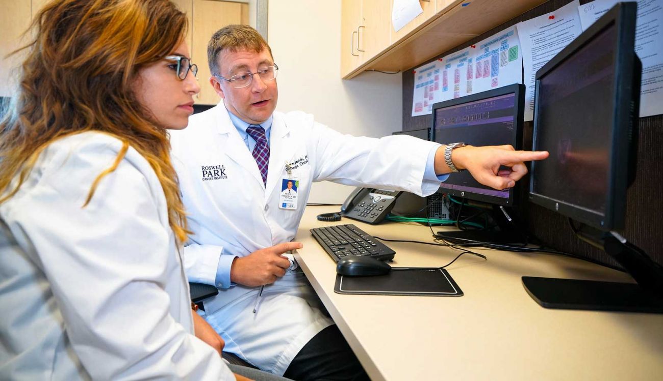
At Roswell Park, we have some of the most advanced imaging tools and pathology resources available so that we get your diagnosis right from the start.
Not all cancers are created equal. Each is comprised of cells that vary from cancer to cancer, and from individual to individual. And tumors may range in size, from millimeters to inches. Accurately identifying the extent of disease and the makeup of your cancer is imperative when determining the best possible approach to treatment.
If you have a symptom that suggests vulvar cancer, your doctor may check general signs of health and may perform the following:
- Pelvic exam: The doctor checks the vagina, uterus, bladder and rectum for any irregularities. To see the upper part of the vagina and the cervix, the doctor inserts an instrument called a speculum into the vagina.
- Biopsy: The doctor removes a sample of tissue from the suspicious area.
- If the abnormal area is small, it may be completely removed by an excisional biopsy. In this procedure, the doctor uses a scalpel to remove a small ellipse of skin and sometimes sews the skin edges together with surgical thread.
- If the abnormal area is larger, a punch biopsy is used to take a small sample. No stitches are needed after the punch biopsy. A pathologist will look at the tissue sample under a microscope to see if cancer or a pre-cancerous condition is present and, if so, what type it is.
State-of-the-art imaging technology
With some of the most advanced imaging tools at our fingertips, and physicians trained to maximize their potential, we consistently provide reliable diagnostic results. Quality imaging enables the Roswell Park medical team to develop the best treatment plan and helps the surgeon map the most direct and effective approach to remove an identified tumor.
One of the more revolutionary imaging devices, the combined Positron Emission Tomography (PET)/Computed Tomography (CT) scanner is currently used to give a total-body overview of glucose (sugar) metabolism, which can reveal metabolic changes of cancer before anatomic abnormalities can be detected with conventional imaging tools such as stand-alone CT and ultrasound.
Special software is used to fuse PET images with CT scans, providing a union that is both functional and anatomic. PET-CT scans can differentiate malignant from benign tissue and can lead to early detection of recurring cancer. They can also grade tumors, define distant metastases, assist in treatment selection and evaluation.