What we do
The Translational Imaging Shared Resource (TISR) at Roswell Park Comprehensive Cancer Center provides specialized preclinical imaging services including image acquisition, quantitative image analysis, multidimensional renderings and data sets for research purposes.
Our mission is to:
- Provide the necessary intellectual and physical resources relating to non-invasive in vivo imaging.
- Develop novel preclinical imaging techniques to meet the needs of researchers and provide insight into various biological processes involved in cancer progression.
- Enable the conduct of preclinical trials of experimental therapeutics in preclinical models.
- Establish technology platforms that allow translation of imaging techniques evaluated in preclinical models to the clinical arena.
Imaging technologies
MRI
Optical
Ultrasound/PAI
X-Ray
Magnetic Resonance Imaging
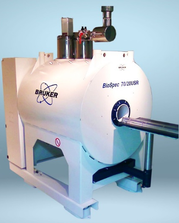
TISR features a state-of-the-art 7T research MRI (Bruker Biospin) that provides full anatomical and functional imaging capabilities.
Capabilities include:
- Anatomical imaging and disease tracking with T1, T2-weighted scans.
- Contrast-enhanced and time of flight angiography.
- Tissue perfusion characterization.
- Localized spectroscopy for relative metabolite concentrations.
- Whole body phenotyping, e.g., body fat composition.
- MR contrast agent relaxometry and in vivo testing.
- Tissue cellularity measurement via water diffusion measurement.
- High-resolution MR microscopy of fixed tissue.
- Customized applications, e.g. MR thermometry and more.
Whole body optical imaging
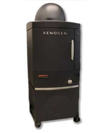
An IVIS Spectrum (Perkin Elmer) offers high-throughput, low-cost screening of disease models.
Capabilities include:
- Monitoring disease states and metastatic burden via luciferase-labeled cells.
- Tracking of fluorescent protein-expressing cells.
- Monitoring gene expression with luciferase reporters.
- Interrogating tissue microenvironment with numerous fluorescent probes.
Ultrasound and photoacoustic imaging
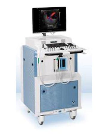
A FujiFilm Vevo 2100/LAZR ultrasound and pulse-laser imaging system.
Capabilities include:
- 3D tissue and tumor volume measurements with calibrated step-motor.
- Tissue hemoglobin levels and oxygenation saturation with photoacoustic system.
- Color doppler for real-time flow rate measurements in vessels.
- Tissue perfusion with ultrasound microbubble contrast.
- Cardiac imaging with artificial intelligence-assisted analysis.
- Power doppler for non-directional flow in tissues.
Additional services
- Bone structural integrity with Faxitron X-ray
- Customized, in-house processing algorithms for high-throughput image analysis
Equipment details
- 7 Tesla, 20 cm bore USR magnet
- ParaVision 360 imaging platform
- MR Imaging Plus and Spectroscopy packages
- Diffusion, DCE, IntraGate cardiac, EPIFlowmap, Short TE, Perfusion, PRESS, STEAM, CSI
- AVANCE NEO electronics with high power, 500 V/ 300A gradient amplifier
- Broadband transmit/receive
- Four-channel parallel imaging
- Coils
- 86 mm(ID) volume coil
- 40 mm (ID) volume coil
- (2) Preclinical, 4-channel brain arrays
- Planar surface coils (10mm, 20mm)
- 1H/19F multi-nuclear coils.
- Physiologic monitoring and gating control
- Thermoelectrically-cooled charge-coupled device (CCD) camera (nominal operating temp -90°C, CCD sensor size: 6.76 cm2
- Four magnification settings capable of imaging fields of view ranging from 3.9 cm x 3.9 cm (20 micron resolution) to 23 cm x 23 cm
- Fluorescence imaging light is provided by a 150W quartz tungsten halogen lamp situated behind a filter wheel comprised of 10 narrow band excitation filters (430-745 nm, 30 nm bandpass).
- 24-position filter wheel featuring 18 emission filters (500-840nm, 20 nm band pass) with full demixing capabilities.
- Linear array with step-motor for 3D reconstruction (5.2 x 10⁻⁵ cu mm voxel size)
- Nd:YAG laser with a tunable range of 680-950 nm for photoacoustic imaging.
- Acquisition Packages:
- 3D mode
- Color, power, PW doppler
- Non-linear contrast modes
- PA-NanoStepper
- PA-Oxyhemo
- Transducers
- LZ 250 (21 MHz, 23x20 mm FOV)
- MS-55 (22-55 MHz, 12x11 mm FOV)
- LZ-201 (9-18 MHz, 32x30 mm FOV)
- Faxitron MX-20 radiographic imaging system
- Adjustable dose for desired contrast
- 20 lines/mm resolution
Software packages
- Multimodal medical imaging data analysis
- Full-array of image data analysis
- 3D visualization
- Rapid data segmentation and ROI sampling
- Multidimensional filtering
- 2D/3D co-registration
- Commonly used platform for developing custom image analysis routines
- Over 100 custom-designed algorithms for efficient data analysis and reporting
- 11 MATLAB Toolboxes including image processing and statistics and machine learning
- Multiple copies located onsite for off-line analysis of IVIS Spectrum data
- Training video available for efficient analysis
- Remote analysis via Citrix Workstation
- Offline analysis via dongle
- Analysis modules include:
- Vevo Multiplexer
- PA-Mode Plus
- 3D Thresholding
- Vevo CQ analysis (Perfusion)
- Auto LV Analysis (Cardiac)
- LV Analysis (Cardiac)
- Free, open-source available
- Medical image analysis. Includes registration, interactive segmentation, visualization and volume rendering
- Many additional extensions available
- 2D/3D quantification
- 3D visualization & animation
Meet our team

Drs. Seshadri and Spernyak jointly address the research needs of investigators and provide their scientific expertise to enable the conduct of imaging research studies in a timely and cost-effective manner.
Services & fees
Please contact either Dr. Joseph Spernyak (716-845-1551, Joseph.Spernyak@RoswellPark.org) or Dr. Mukund Seshadri (716-845-1552, Mukund.Seshadri@RoswellPark.org) for a project-specific cost estimate.
Information for publications
All presentations/posters/publications utilizing data acquired from TISR equipment and/or input from TISR staff must acknowledge Roswell Park’s Comprehensive Cancer Center Support Grant: P30CA016056.
Additionally, the appropriate shared instrumentation (S10) grant must also be acknowledged if data was acquired on the following equipment:
- 7T MRI: S10OD030397
- IVIS Spectrum: S10OD16450
- Vevo LAZR/Ultrasound: S10OD10393
Useful links
FOM Reservation calendar Perkin-Elmer IVIS training videos (YouTube)
Analyze 14 tutorials (YouTube)Visualsonics tips and tricks
Location and hours
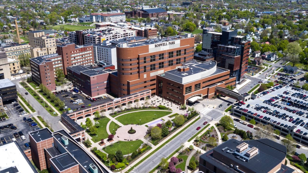
Roswell Park Comprehensive Cancer CenterTranslational Imaging Shared Resource
Cancer Cell Center, Room 164
Elm and Carlton Streets
Buffalo, New York 14263
Operating schedule
Monday – Friday, 8 a.m. – 5 p.m. (Staffing Present)
TISR provides 24/7 access to imaging technologies to fully trained staff of Cancer Center Support Grant (CCSG) members’ laboratories.
Publications
Clinical Cancer Research, Seshadri et al., 2005

Journal of Biological Chemistry, Jell et al., 2007

Journal of Cerebral Blood Flow & Metabolism, Seshadri and Ciesielski., 2009

Angiogenesis, Seshadri et al., 2011

Oral Oncology, Folaron et al., 2013

Cancer Prevention Research, Bothwell et al., 2015

Inorganic Chemistry, Olatunde, et al. 2014

Journal of Oral Pathology & Medicine, Verma, et.al. 2018
- Cheruku RR, Turowski SG, Durrani FA, Tabaczynski WA, Cacaccio J, Missert JR, Curtin L, Sexton S, Alberico R, Hendler CM, Spernyak JA, Grossman Z, Pandey RK. Tumor-Avid 3-(1'-Hexyloxy)ethyl-3-devinylpyrpyropheophorbide-a (HPPH)-3Gd(III)tetraxetan (DOTA) Conjugate Defines Primary Tumors and Metastases. J Med Chem. 2022 Jul 14;65(13):9267-9280 PMID: 35763292.
- Kumar R, Chaudhary AK, Woytash J, Inigo JR, Gokhale AA, Bshara W, Attwood K, Wang J, Spernyak JA, Rath E, Yadav N, Haller D, Goodrich DW, Tang DG, Chandra D. A mitochondrial unfolded protein response inhibitor suppresses prostate cancer growth in mice via HSP60. J Clin Invest. 2022 Jul 1;132(13):e149906. PMID: 35653190; PMCID: PMC9246382.
- Sokolow GE, Crawley MR, Morphet DR, Asik D, Spernyak JA, McGray AJR, Cook TR, Morrow JR. Metal-Organic Polyhedron with Four Fe(III) Centers Producing Enhanced T1 Magnetic Resonance Imaging Contrast in Tumors. Inorg Chem. 2022 Feb 7;61(5):2603-2611. PMID: 35073060; PMCID: PMC9038074.
- DeJohn CR, Grant SR, Seshadri M. Application of Machine Learning Methods to Improve the Performance of Ultrasound in Head and Neck Oncology: A Literature Review. Cancers (Basel). 2022 Jan 28;14(3):665. PMID: 35158932; PMCID: PMC8833587.
- Vincent-Chong VK, Seshadri M. Adrenergic-Angiogenic Crosstalk in Head and Neck Cancer: Mechanisms and Therapeutic Implications. Front Oral Health. 2021;2. PMID: 34790909; PMCID:PMC8594278.
- Sonkawade SD, Pokharel S, Karthikeyan B, Kim M, Xu S, Kc K, Sexton S, Catalfamo K, Spernyak JA, Sharma UC. Small Endogeneous Peptide Mitigates Myocardial Remodeling in a Mouse Model of Cardioselective Galectin-3 Overexpression. Circ Heart Fail. 2021:Circheartfailure121008510. PMID: 34415177.
- Panta P, Dhopathi SR, Gilligan G, Seshadri M. Invasive oral squamous cell carcinoma induced by concurrent smokeless tobacco and creamy snuff use: A case report. Oral Oncol. 2021:105354. PMID: 34023217.
- Momeni A, Eagler L, Lo CY, Weil BR, Canty JM, Jr., Lang JK, Neelamegham S. Neutrophils aid cellular therapeutics by enhancing glycoengineered stem cell recruitment and retention at sites of inflammation. Biomaterials. 2021;276:121048. PMID: 34343858.
- Liu Z, Seshadri M, Gupta V, Papanicolau-Sengos A, Merzianu M. INI1-Deficient Thyroid Carcinoma is an Aggressive Disease with Epithelioid and Rhabdoid Phenotype. A Case Report, Survey of INI1 Expression in Thyroid Lesions and Literature Review. Head Neck Pathol. 2021. PMID: 34057693.
- Kras EA, Abozeid SM, Eduardo W, Spernyak JA, Morrow JR. Comparison of phosphonate, hydroxypropyl and carboxylate pendants in Fe(III) macrocyclic complexes as MRI contrast agents. J Inorg Biochem. 2021;225:111594. PMID: 34517167.
- Knudsen ES, Kumarasamy V, Chung S, Ruiz A, Vail P, Tzetzo S, Wu J, Nambiar R, Sivinski J, Chauhan SS, Seshadri M, Abrams SI, Wang J, Witkiewicz AK. Targeting dual signalling pathways in concert with immune checkpoints for the treatment of pancreatic cancer. Gut. 2021;70(1):127-38. PMID: 32424005; PMCID:PMC7671951.
- King G, Veros KM, MacLaren DAA, Leigh MPK, Spernyak JA, Clark SD. Human wildtype tau expression in cholinergic pedunculopontine tegmental neurons is sufficient to produce PSP-like behavioural deficits and neuropathology. Eur J Neurosci. 2021;54(10):7688-709. PMID: 34668254.
- Dykstra KM, Fay HRS, Massey AC, Yang N, Johnson M, Portwood S, Guzman ML, Wang ES. Inhibiting autophagy targets human leukemic stem cells and hypoxic AML blasts by disrupting mitochondrial homeostasis. Blood Adv. 2021;5(8):2087-100. PMID: 33877295; PMCID:PMC8095145.
- Damasco JA, Ohulchanskyy TY, Mahajan S, Chen G, Singh A, Kutscher HL, Huang H, Turowski SG, Spernyak JA, Singh AK, Lovell JF, Seshadri M, Prasad PN. Excretable, ultrasmall hexagonal NaGdF4:Yb50% nanoparticles for bimodal imaging and radiosensitization. Cancer Nanotechnol. 2021;12(1):4. PMID: 33603920; PMCID:PMC7864820.
- Asik D, Abozeid SM, Turowski SG, Spernyak JA, Morrow JR. Dinuclear Fe(III) Hydroxypropyl-Appended Macrocyclic Complexes as MRI Probes. Inorg Chem. 2021;60(12):8651-64. PMID: 34110140.
- Anandakrishnan N, Ye H, Guo Z, Chen Z, Mentkowski KI, Lang JK, Rajabian N, Andreadis ST, Ma Z, Spernyak JA, Lovell JF, Wang D, Xia J, Zhou C, Zhao R. Fast Stereolithography Printing of Large-Scale Biocompatible Hydrogel Models. Adv Healthc Mater. 2021:e2002103. PMID: 33586366.
- Abozeid SM, Chowdhury MSI, Asik D, Spernyak JA, Morrow JR. Liposomal Fe(III) Macrocyclic Complexes with Hydroxypropyl Pendants as MRI Probes. ACS Appl Bio Mater. 2021;4(11):7951-60. PMID: 35006776.
This resource is funded by the Roswell Park Comprehensive Cancer Center Support Grant (CCSG) awarded by the National Cancer Institute (NCI P30CA16056). Publications that have utilized facility resources, services or scientific data generated by the resource should acknowledge the resource or the assistance provided by resource staff and cite the NCI CCSG grant. An electronic copy of the publication should also be sent to the resource directors.
Additionally, please acknowledge NOH Shared Instrumentation Grants if data as acquired from the 7T MRI (S10OD030397) IVIS Spectrum (S10OD16450) or Vevo LAZR ultrasound/photoacoustic system (S10OD10393)
Sample language
The work utilized resources and services provided by the Translational Imaging Shared Resource at Roswell Park are supported by NCI P30CA16056. The authors would also like to acknowledge the resource staff for their technical assistance in performing the imaging studies.
The Translational Imaging Shared Resource (TISR) at Roswell Park Comprehensive Cancer Center provides scientific and technical services pertaining to multimodality image acquisition, quantitative image analysis, multidimensional renderings and visualization of imaging data sets.
The resource is equipped with (a) a Bruker 7.0 Tesla MRI, (b) a preclinical ultrasound/photoacoustic imaging system (Vevo® LAZR, VisualSonics), (c) a combined bioluminescence/fluorescence imaging system (IVIS Spectrum, Perkin-Elmer) with tomographic and multi-spectral capabilities, (d) a bioluminescence imaging system (IVIS-50, Perkin-Elmer), and (e) a planar X-RAY imaging system (MX-20, Faxitron). The resource offers a wide range of anatomical and functional imaging services including disease monitoring, tumor microvascular/ oxygenation assessment and novel imaging agent development. Resource staff also facilitate multimodality imaging research through collaborative interactions with other core resources or regionally available instrumentation (e.g., microPET imaging, State University of New York at Buffalo, Department of Nuclear Medicine).