As technology advances, it provides new options for diagnosing gastrointestinal (GI) conditions.
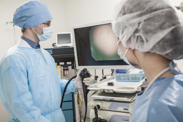
High-definition cameras are used to view the GI tract in a variety of ways; upper endoscopies and colonoscopies are routine procedures that help a doctor view the inside of the esophagus, stomach and colon. A laparoscopy can be performed to view the outside of the bowel with a small incision made in the abdomen.
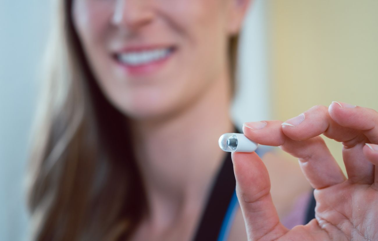
But how would they view the area in between your stomach and colon — those 20 feet of small intestines — without an incision or even sedation? This is where a capsule endoscopy comes in.
“A capsule endoscopy is a very small camera the patient swallows that takes a series of pictures as it travels through the intestines,” says gastroenterologist Kevin Robillard, MD, Associate Professor of Oncology in the Department of Internal Medicine at Roswell Park Comprehensive Cancer Center. “Video endoscopies are performed to look at areas that are hard to reach with a standard endoscopy, given the length of the small intestines. It gives us the ability to look for such things as tumors, polyps, damage to the intestines or bleeding lesions.”
“We’ll turn it on before the patient swallows it so it can start recording immediately and begin sending the information to a recorder that the patient is wearing.”
This recorder is a small device the patient wears on a belt throughout the day, wirelessly receiving and downloading the photos the capsule is capturing. “The capsule will record images until the battery runs out,” explains Dr. Robillard. “It records, currently, for up to twelve hours. Once the battery runs out, a light on the recorder will change color, and the recording is done.” It’s then that the patient will return the recording device to their doctor, where the images will be downloaded and reviewed.
“What I typically do first is check the end of the video, to make sure the capsule visualizes the colon,” explains Dr. Robillard. If the video footage ends in the colon, this is evidence that the capsule has been — or soon will be — passed in a bowel movement. “If in the video footage we don’t see it enter the colon, then we’d let the patient know to keep an eye out for it.” Once it has passed, the patient is not required to retrieve or return the capsule. An X-ray will sometimes be used to confirm the passage.
Never miss another Cancer Talk blog!
Sign up to receive our weekly Cancer Talk e-newsletter.
Sign up!Prep for a capsule endoscopy
Preparation for a capsule endoscopy is minimal. “Patients fast, nothing to eat or drink, for 12 hours prior to the procedure. We ask patients to hold off on taking their medications immediately prior to swallowing the capsule, because some medications have coloring in them,” says Dr. Robillard. “If you fast prior to the procedure, it is unlikely to have food residue remaining in the GI tract. If it enters the small intestine and there is some residue, we may not be able to see well.”
Similarly, conditions like gastroparesis, or slower stomach-emptying could lead to the capsule being in the stomach for the majority of the battery life – meaning little to no images are captured of the small intestine, which is the main goal of the procedure.
To limit the risk of residue and blockage of the camera, there is a four-hour fasting period after the capsule is swallowed. “We don’t want to see a hamburger floating past the camera,” Dr. Robillard jokes. Four hours after swallowing the capsule, patients are permitted to have a light meal and clear liquids.
The majority of the time, no bowel prep is required leading up to a capsule endoscopy. “Usually, we will only do prep if there has been a capsule endoscopy done in the past where the visualization was suboptimal,” says Dr. Robillard. In these cases, Dr. Robillard may perform an upper endoscopy to manually place the capsule in the small intestine.
The risk and benefits
Because the main purpose of performing a capsule endoscopy is to get a view of the small intestines, there are many benefits to this non-invasive procedure. “As long as it’s swallowed effectively, the procedure is extremely safe,” Dr. Robillard reassures his patients. “There is no sedation involved, so the patient can drive themselves in and home.”
“The only real risk is that the capsule could get stuck at a narrow point,” says Dr. Robillard. “If it does get stuck, that’s abnormal. But, that would mean a lot of things are getting stuck there, and that would help diagnose a problem we’re looking for.” In such a situation, a double balloon endoscopy could be performed to both remove the capsule and look further into the issue that the capsule has found. Alternative options to treat such a condition could be medications, like a steroid, to help open up the narrow point, or surgery to eliminate the narrowing.
“You can get a tumor of the small intestines either because the tumor started there, or it ended up there because of metastatic disease,” says Dr. Robillard. “When we suspect something may be cancer-related, Roswell Park is where you want to have this imaging done.”